44 sea urchin anatomy diagram
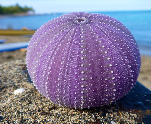
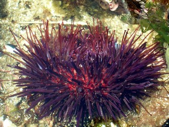
Sea Urchin Anatomy One look at a sea urchin and you can see why they would be called sea hedgehogs. They have hard rounded shells covered with sharp movable spines. Urchins are part of the phylum Echinoderm and their name comes from Ancient Greek (echinos meaning "hedgehog" and derma meaning "skin"). There are more than 900 species of sea urchins and they come in a range of What is a sea urchin? A sea urchin is a small marine animal in the Echinodermata phylum that is spherical in shape and covered in spines or cilia. There are 950 species of sea urchins. Some are irregular, meaning their appearance or anatomy varies from the majority of the species. Where do sea urchins live?
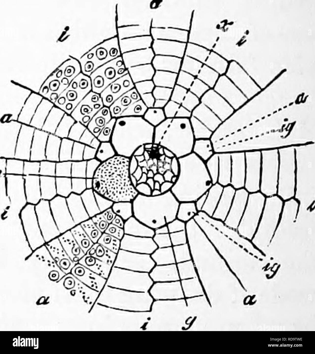
This is the rudiment and it is these cells that give rise to the adult sea urchin. As the rudiment develops, adult features, such as tube-feet, begin to appear, while larval structures, including the arms and even the larval gut and mouth, are resorbed and eventually lost. ... (inner cycle in diagram).

Sea urchin anatomy diagram
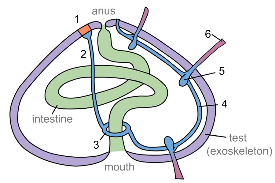
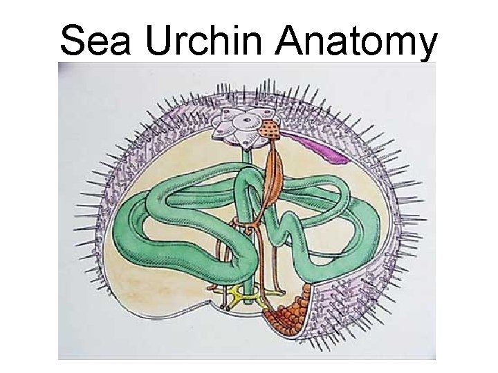
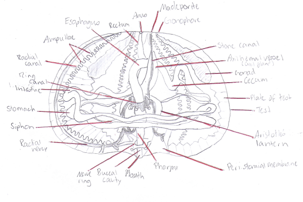
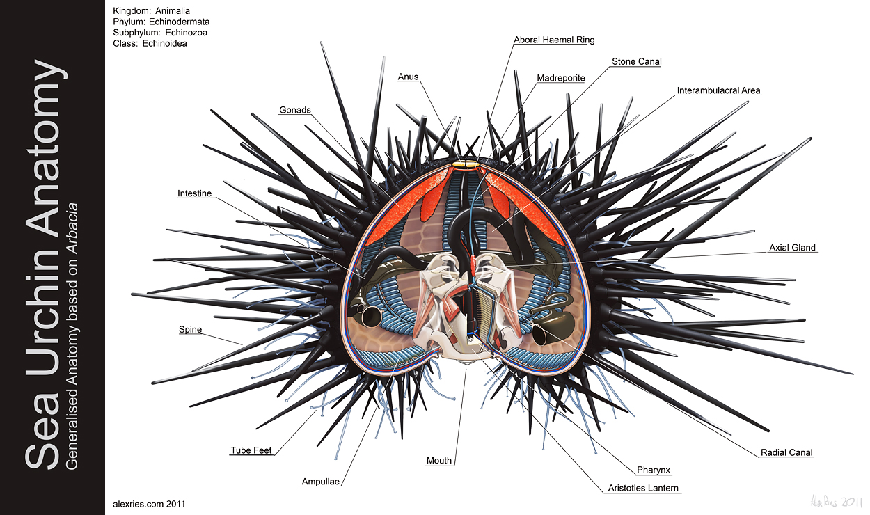
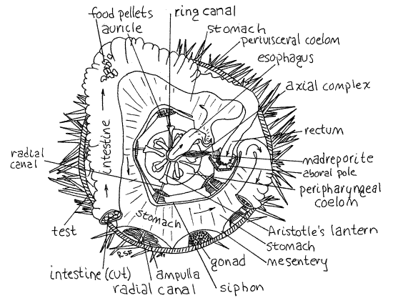
Anatomy chapter 6 quizlet. Start studying anatomy chapter 6. Bones and skeletal tissue flashcards at proprofs these flashcards are for chapter 6. Learn vocabulary terms and more with flashcards games and other study tools. Anatomy and physiology chapter 6 part a. Layer of dense irregular connective tissue. Download scientific diagram | The anatomy of the sea urchin Strongylocentrotus droebachiensis. from publication: A Guide to the Sea Urchin Reproductive Cycle and Staging Sea Urchin Gonad Samples ... Sea Urchin Anatomy In Detail. Sea Urchin Anatomy In Detail. In this image, you will find gonads, anus, aboral haemal ring, madreporite, stone canal, interambulacral area, axial gland, radial canal, pharynx, Aristotle’s lantern, öouth, ampullae, tube feet, spine, intestine in it. Health care advices from Overseas Doctor .
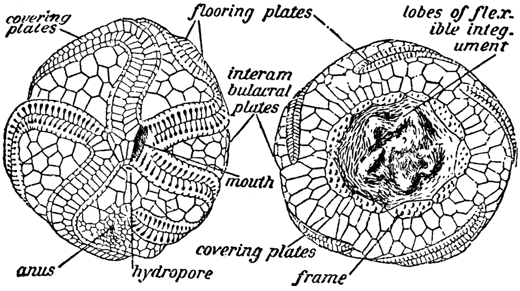
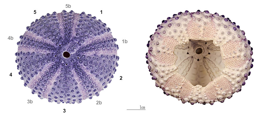
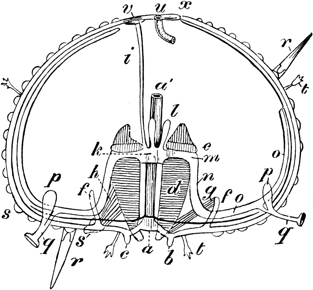
Sea urchin anatomy diagram. In this article we will discuss about the structure of Sea Urchin (Echinus) with the help of a diagram. 1. It is commonly known as “sea urchin” and is formed in shallow water in both rocky and sandy place in sea. 2. Body is Sub-globular and convex or dome-shaped above and flattened below. The aboral and oral surfaces are distinct. 3 ... Sea urchins (/ ˈ ɜːr tʃ ɪ n z /) are spiny, globular echinoderms in the class Echinoidea.About 950 species of sea urchin live on the seabed of every ocean and inhabit every depth zone — from the intertidal seashore down to 5,000 metres (16,000 ft; 2,700 fathoms). Sea Urchin Dissections 1. Wear new latex gloves at all times a. The human hands contain RNA degrading enzymes that can digest delicate RNA molecules 2. Pre-label the cryovials with a. Date of dissection b. Urchin genus and species name c. Collection location d. Tissue type (example: digestive, intestine, stomach, etc) e. ID number (example ... Question 41. SURVEY. 30 seconds. Q. The diagram represents reproduction and cell division in a sea urchin (animal) in stages labeled A through F. When a sea urchin reproduces, the female sex cell and the male sex cell unite to form a cell called a zygote. The zygote divides several times in a matter of hours.
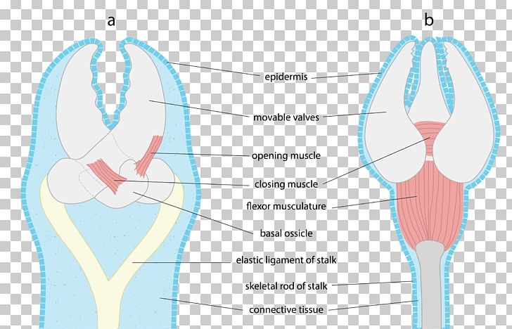
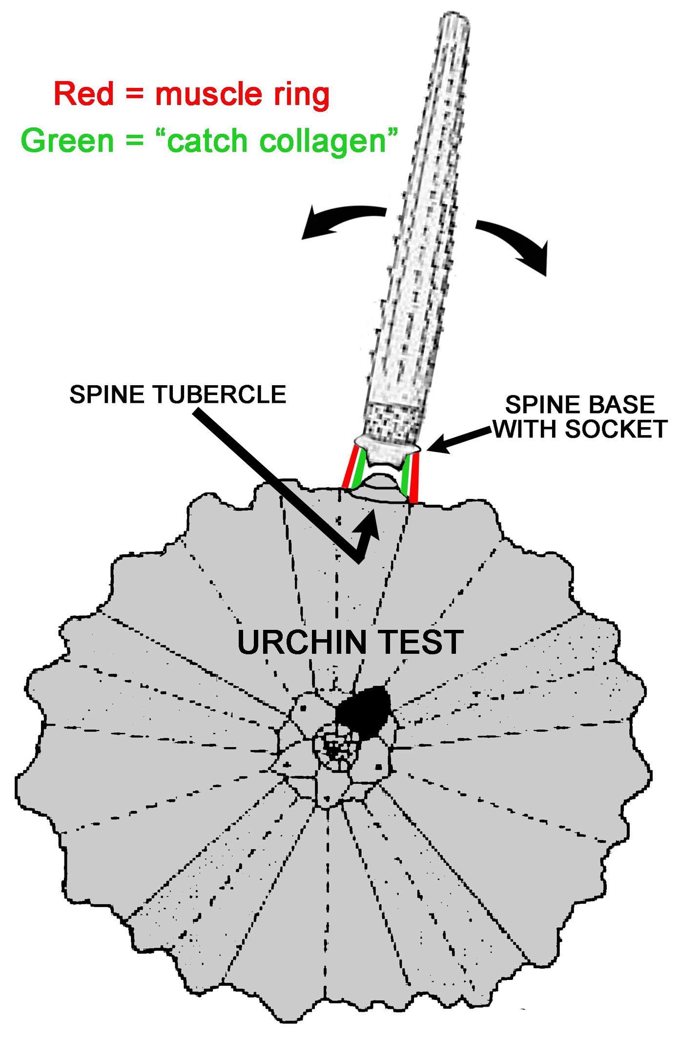
Sea urchins have no "brain" as we think of it, instead they have a nerve ring near the hydraulic system used to power the tube feet. THE TUBE FEET. The tube feet are the primary means of locomotion for the sea urchin (along with moving the spines). Lateral movement is accomplished by means of paired muscles in the tube foot itself and extension and retraction by muscles in the hydraulic "bulb" behind each tube foot. While each "foot" may seems weak and ineffective, the large numbers of feet ... Leg legs are the lower angled strokes which you can see in the letters k r and q. ... Frog Anatomy Diagrams 15.54 Zion Clark Anatomy 12.49 The Anatomy Of A Synapse Answer Key 17.08 Sea Urchin Anatomy 00.18 Microscopic Anatomy And Organization Of Skeletal Muscle 22.07 Anatomy Drawing References 00.08 Female Anatomy Nude 03.55 Sea Urchin Anatomy In Detail. Sea Urchin Anatomy In Detail. In this image, you will find gonads, anus, aboral haemal ring, madreporite, stone canal, interambulacral area, axial gland, radial canal, pharynx, Aristotle’s lantern, öouth, ampullae, tube feet, spine, intestine in it. Health care advices from Overseas Doctor . Download scientific diagram | The anatomy of the sea urchin Strongylocentrotus droebachiensis. from publication: A Guide to the Sea Urchin Reproductive Cycle and Staging Sea Urchin Gonad Samples ...
Anatomy chapter 6 quizlet. Start studying anatomy chapter 6. Bones and skeletal tissue flashcards at proprofs these flashcards are for chapter 6. Learn vocabulary terms and more with flashcards games and other study tools. Anatomy and physiology chapter 6 part a. Layer of dense irregular connective tissue.


0 Response to "44 sea urchin anatomy diagram"
Post a Comment