45 diagram of neck muscles
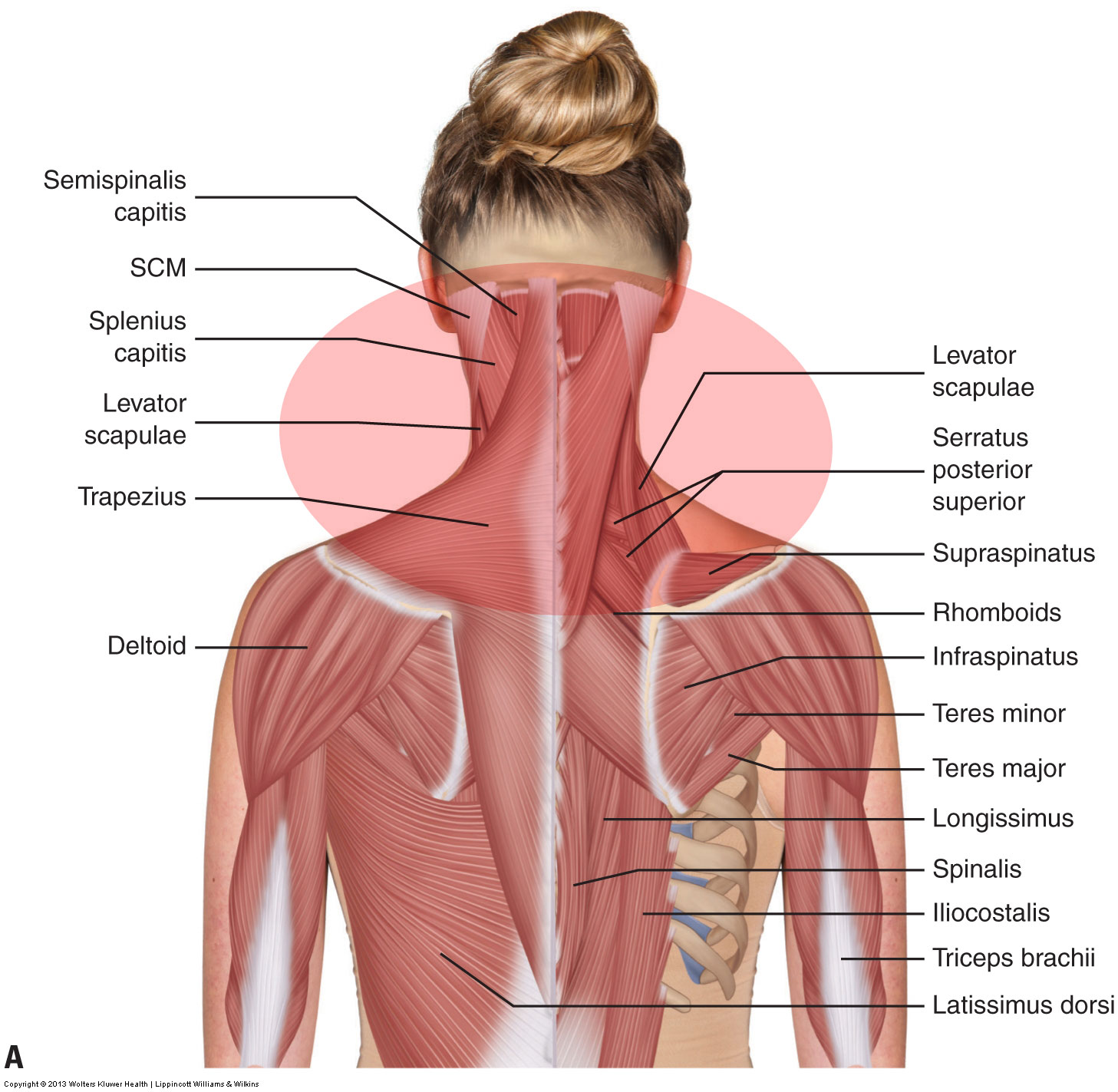
When people talk about their neck muscles it is usually their traps that they are referring to. Your trapezius is a flat, triangle-shaped muscle that extends from your neck, down along your spine down to the middle of your back and across your shoulder blade. There are both right and left traps, and they are used to support your arms and shoulders. Instant anatomy is a specialised web site for you to learn all about human anatomy of the body with diagrams, podcasts and revision questions
Nov 11, 2021 · Back And Neck Muscles Diagram. Health care advices from Overseas Doctor . We are pleased to provide you with the picture named Back And Neck Muscles Diagram. We hope this picture Back And Neck Muscles Diagram can help you study and research. for more anatomy content please follow us and visit our website: www.anatomynote.com.
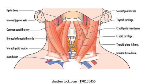
Diagram of neck muscles
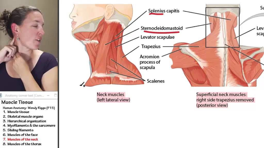
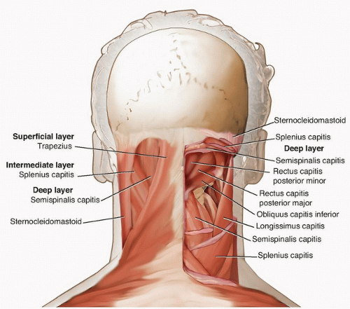
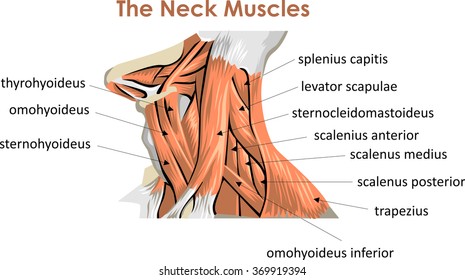
Feb 17, 2015 · Superficial muscles are the muscles closest to the skin surface and can usually be seen while a body is performing actions. Many in the neck help to stabilize or move the head. Some also create ... The muscles of the neck are present in four main groups. The suboccipital muscles act to rotate the head and extend the neck.Rectus capitis posterior major and Rectus capitis posterior minor attach the inferior nuchal line of the occiput to the C2 and C1 vertebrae respectively.Obliquus capitis superior also extends from the occiput to C1 while obliquus capitis inferior originates from C2 and ... The neck muscles, including the sternocleidomastoid and the trapezius, are responsible for the gross motor movement in the muscular system of the head and neck. They move the head in every direction, pulling the skull and jaw towards the shoulders, spine, and scapula. Working in pairs on the left and right sides of the body, these muscles ...
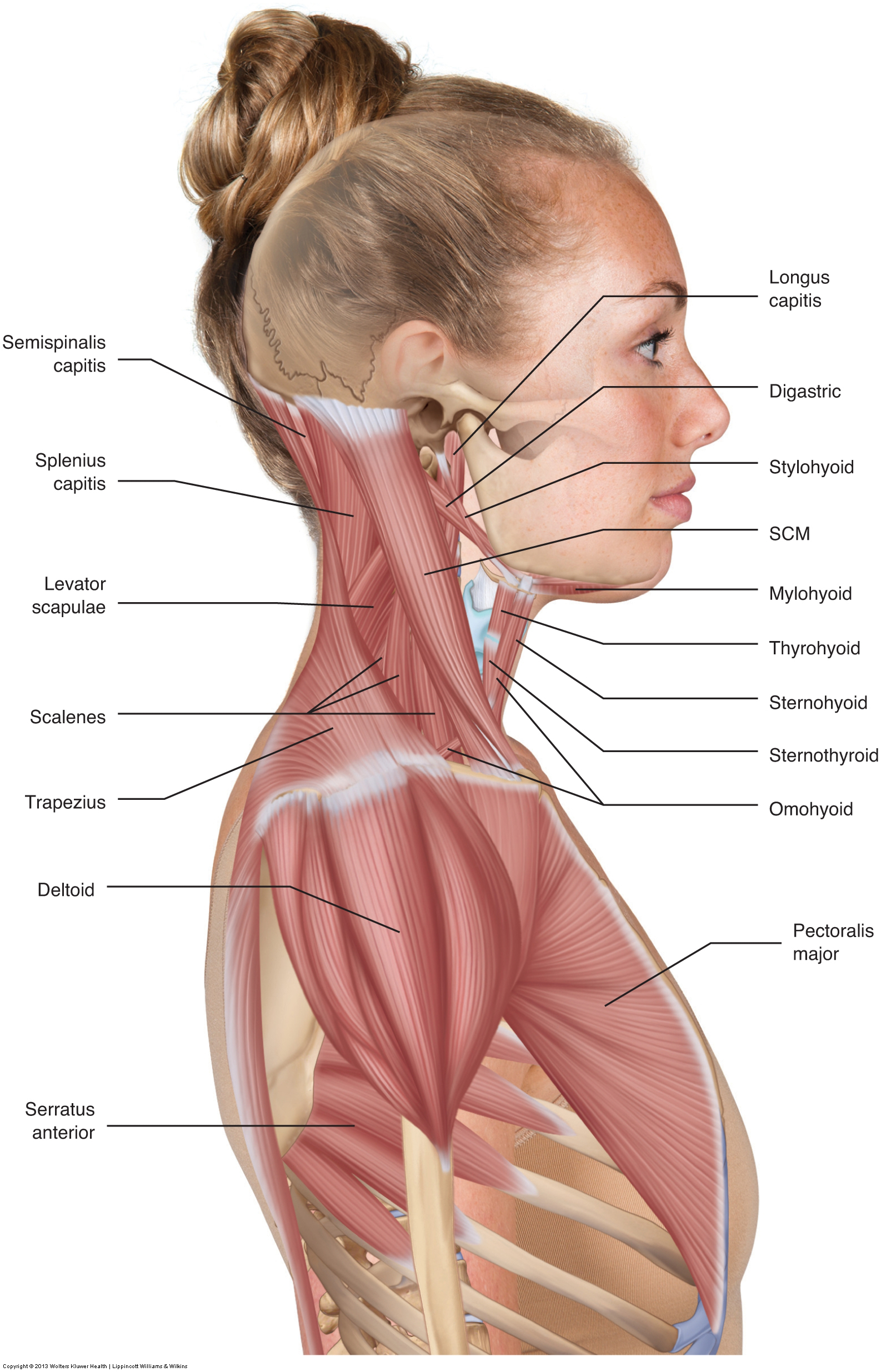
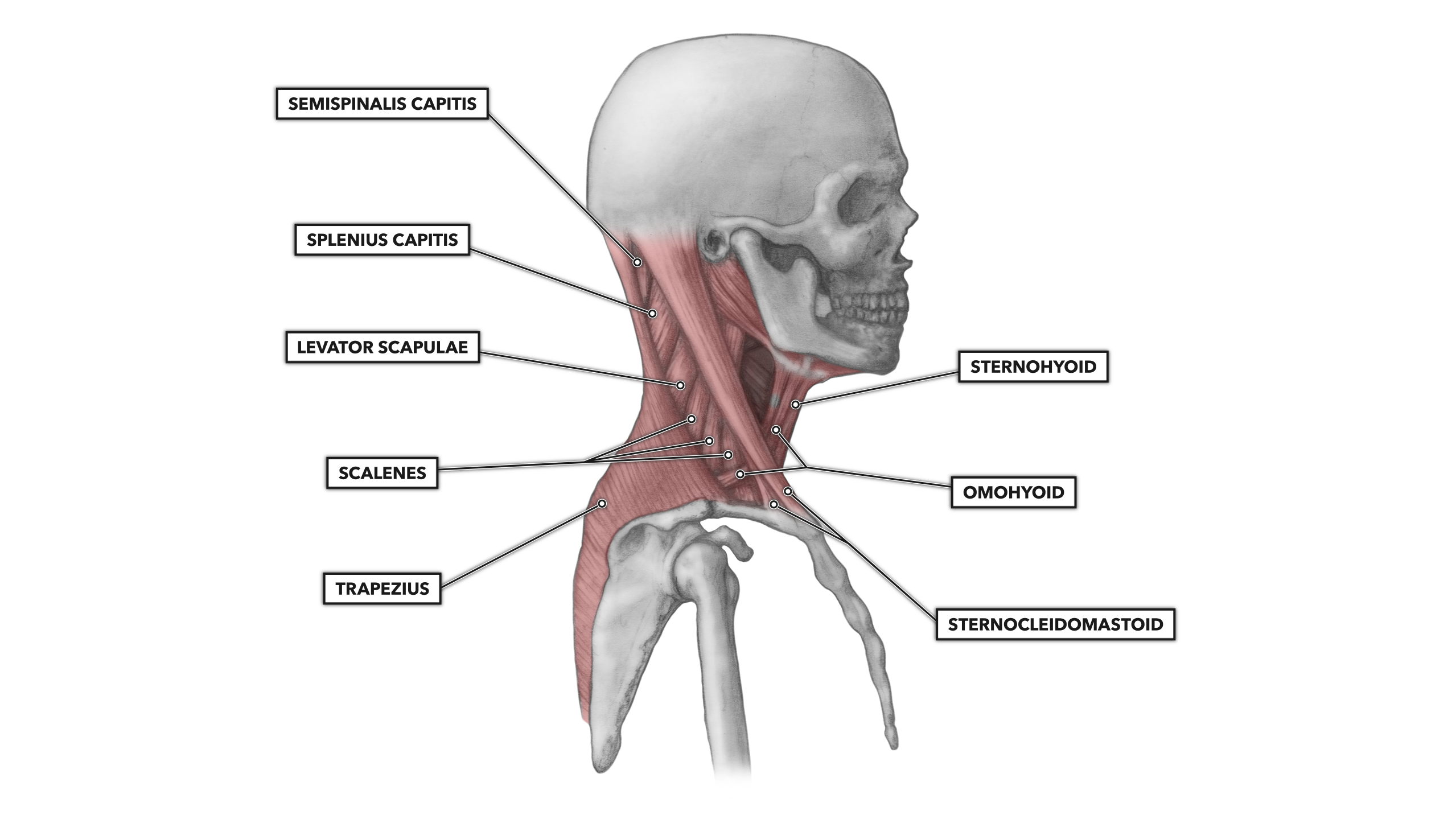
Diagram of neck muscles. The following diagram below is the human body muscle diagram. Check out and click on the image to download it. The muscular system is responsible for the movement of the human body. Attached to the bones of the skeletal system are about 700 named muscles that make up roughly half of a person's body weight. Each of these muscles is a discrete ... The neck is the bridge between the head and the rest of the body. It is located in between the mandible and the clavicle, connecting the head directly to the torso, and contains numerous vital structures. It contains some of the most complex and intricate anatomy in the body and is comprised of numerous organs and tissues with essential structure and function for normal physiology. Labeled Anatomy Chart of Neck and Back Muscles on White Background Labeled human anatomy diagram of man's neck and back muscles from a posterior view on a white background. anatomy of neck and shoulder stock pictures, royalty-free photos & images. May 06, 2015 · Here is a list of the many muscles that exist in the neck. Longus Colli & Capitis – Responsible for flexion of the head and neck. Rectus Capitis Anterior– Responsible for flexion of the neck. Rectus Capitis Lateralis– Helps the neck to bend to the side. Scalene Muscles – Responsible for lifting the first and second ribs i.e. assist with breathing
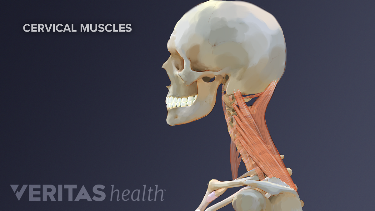
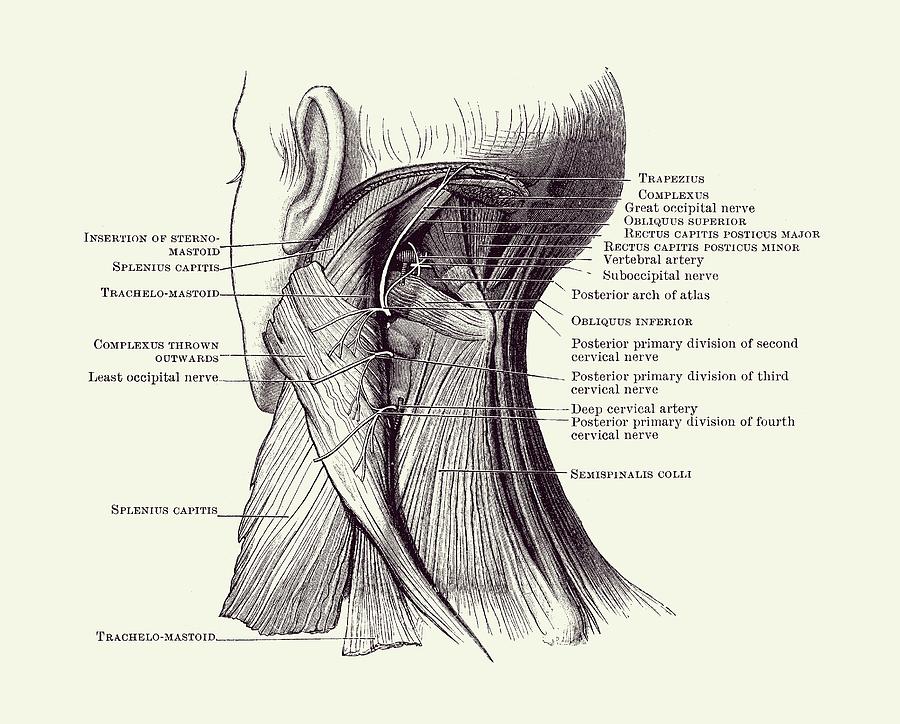
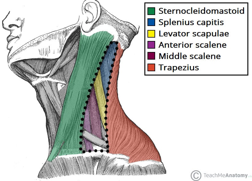
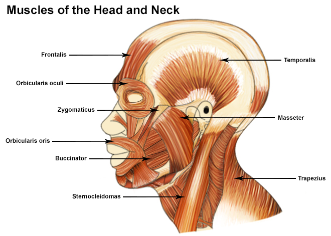
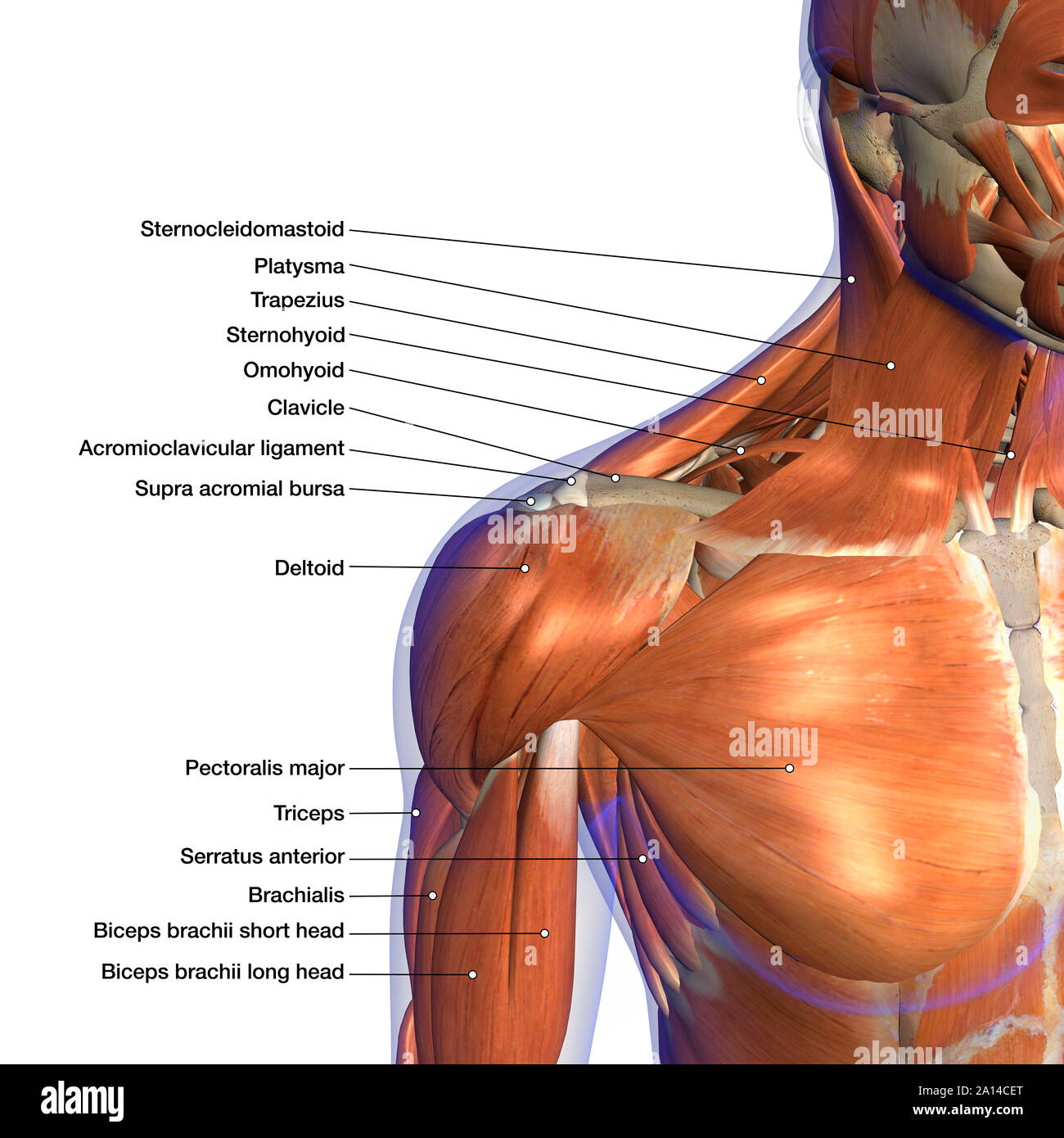
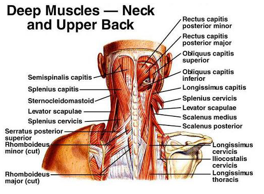
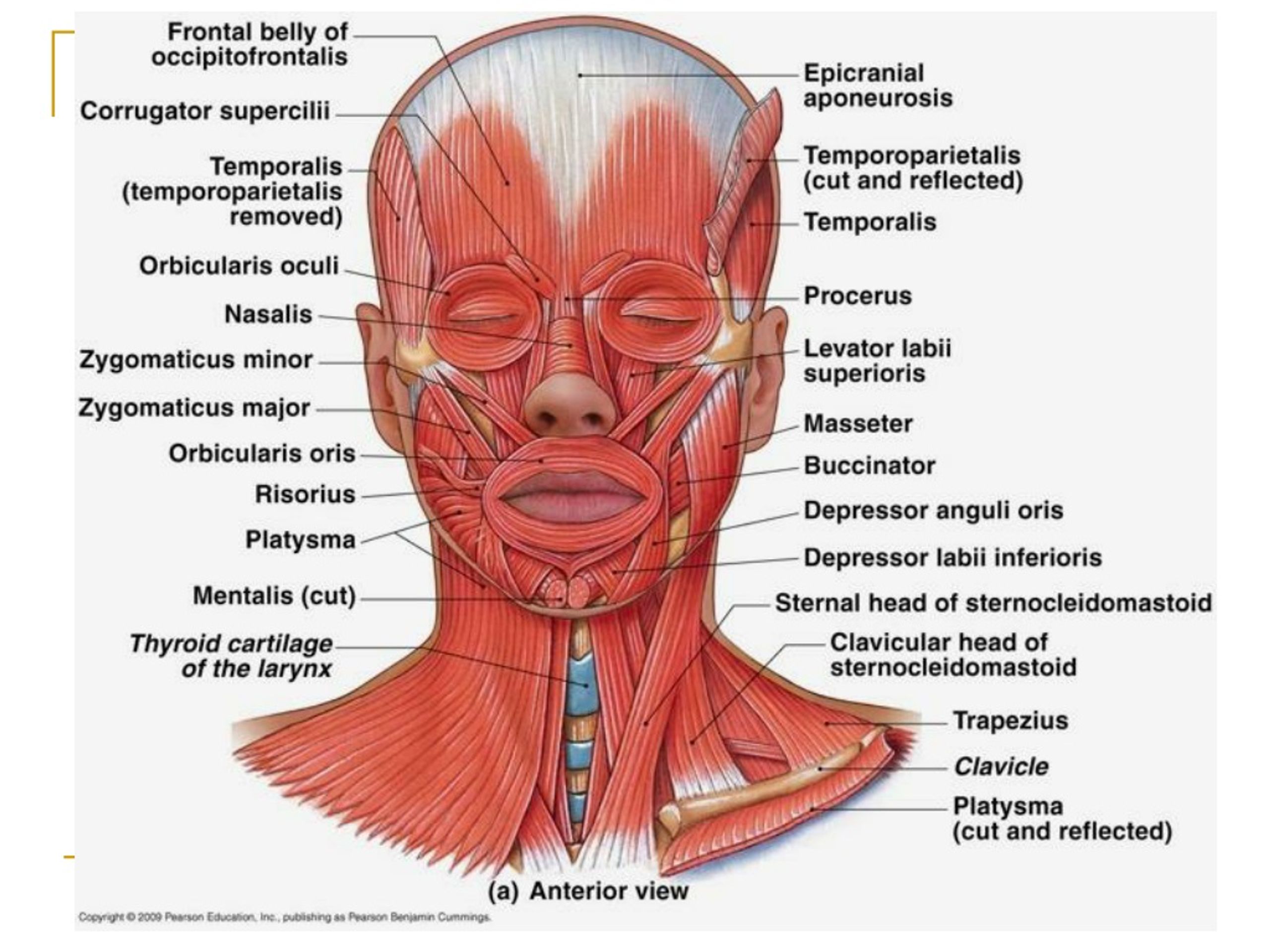
Sep 03, 2019 · Head And Neck Muscles Diagram. In this image, you will find cranial aponeurosis, temporalis, occipitalis, masseter, sternocleidomastoid, trapezius, platysma, orbicularis oris, buccinator, zygomaticus, orbicularis oculi, frontalis in Head and neck muscles diagram. Our LATEST youtube film is ready to run. The Muscles of the Neck anatomical chart shows in beautiful detail the many anterior, posterior, inferior and lateral views of every muscle that makes up the matrix of support for our skull and brain. Included are views of the back of the neck, short muscles of the neck, prevertebral muscles, platysma and more. Neck muscles can be strained from poor posture — whether it's leaning over your computer or hunching over your workbench. Osteoarthritis also is a common cause of neck pain. Rarely, neck pain can be a symptom of a more serious problem. Seek medical care if your neck pain is accompanied by numbness or loss of strength in your arms or hands or ... Prominent muscle of the lateral neck-The sternal head of the sternocleidomastoid originates from the manubrium of the sternum and the clavicular head originates from the medial one third of the clavicle-Both heads insert onto the mastoid process of the temporal bone and the superior nuchal line of the occipital bone
Labeled human anatomy diagram of man's neck and back muscles from a posterior view on a white background. 1 credit. Essentials collection. Everyday photos and illustrations, for just 1 credit. $12 for this image. $4 with a 1-month subscription (10 Essentials images for $40) Continue with purchase. Muscle Charts of the Human Body For your reference value these charts show the major superficial and deep muscles of the human body. Superficial and deep anterior muscles of upper body Nov 11, 2021 · Muscles of the neck (Musculi cervicales) The muscles of the neck are muscles that cover the area of the neck. These muscles are mainly responsible for the movement of the head in all directions. They consist of 3 main groups of muscles: anterior, lateral and posterior groups, based on their position in the neck. The musculature of the neck is further divided into more specific groups based on a number of determinants; including depth, precise location and function. Neck and Shoulder Pain Anatomy. The anatomy of the neck and shoulders is very interesting. These critical parts of the upper body are very prone to developing pain because the position of all the bones in the neck and shoulders are completely dependent on the balance and alignment of the muscles and fascia that lash them together and allow for movement between them.
Anterior Neck Muscles Images Stock Photos Vectors Shutterstock
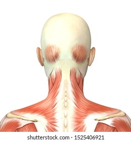
On these diagrams of back muscle, you'll learn about back muscles, their locations and functional anatomy. The latissimus dorsi, also known as the "lats" or "wings," are the largest and most well-known of all the back muscles. The trapezius muscles are located between your shoulder and your neck.
Neck Muscles Images Stock Photos Vectors Shutterstock
For example, some muscles located in the chest also help move the shoulders. Likewise, there are muscles in other parts of the body that help support and move the spine. Below you'll see diagrams along with the names of the back muscles that may be the cause of your pain. How Many Muscles Are in the Back? The back has a total of 40 muscles.
Lymph Node
Jan 20, 2018 · Neck muscles are bodies of tissue that produce motion in the neck when stimulated. The muscles of the neck run from the base of the skull to the upper back and work together to bend the head and ...

Muscles Of The Neck And Trunk Learn Muscles
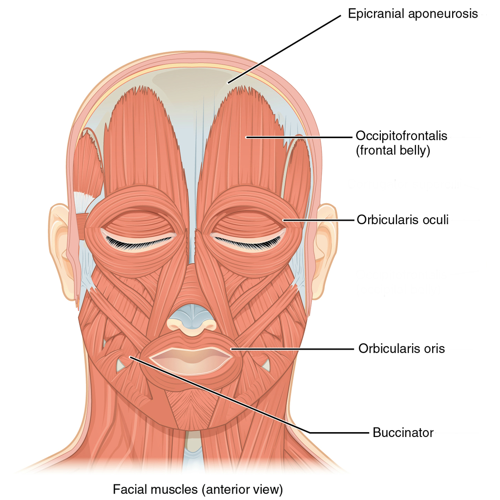
Muscles of the Head and Neck. Humans have well-developed muscles in the face that permit a large variety of facial expressions. Because the muscles are used to show surprise, disgust, anger, fear, and other emotions, they are an important means of nonverbal communication. Muscles of facial expression include frontalis, orbicularis oris, laris ...
Neck Muscles And Other Soft Tissues
Muscles of the Anterior Neck, Chest and Thorax Coloring Page. Muscles of the Arm and Forearm (Anterior) - Coloring Page. ... Nervous System Structure and Function Diagram (Copy Ready) Parts of the Brain Diagram and Coloring Page. Pelvis Boney Features Coloring Page.
Muscles Of The Neck Musculature Of The Cervical Spine
The muscles of the human body can be categorized into a number of groups which include muscles relating to the head and neck, muscles of the torso or trunk, muscles of the upper limbs, and muscles of the lower limbs. The human body has three different types of muscles. They include: skeletal muscles, smooth muscles, cardiac muscles.
Muscle 7 Muscles Of The Neck Youtube
Neck spaces. The content of the neck is grouped into 4 neck spaces, called the compartments.. Vertebral compartment: contains cervical vertebrae and postural muscles.; Visceral compartment: contains glands (thyroid, parathyroid, and thymus), the larynx, pharynx and trachea.; Two vascular compartments: contain the common carotid artery, internal jugular vein and the vagus nerve, on each side of ...
Neck Muscular System Diagram Vintage Anatomy 2 Drawing By Vintage Anatomy Prints
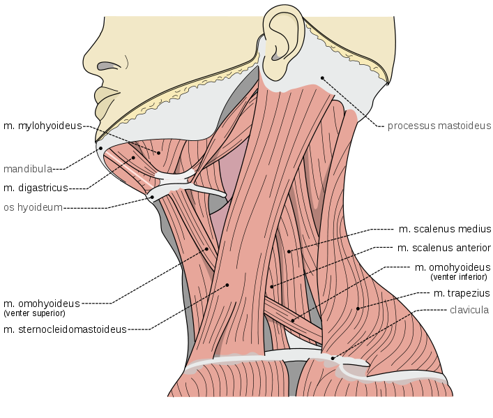
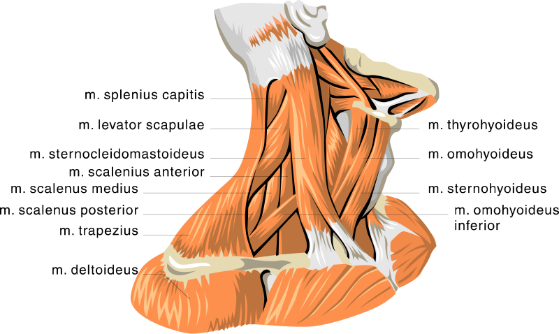
The cervical vertebrae serve as the origination and insertion points for a host of muscles that support but also enable movement of the head and neck. The musculature of the neck is comprised of a number of different muscle groups. They can be divided into anterior, lateral and posterior groups based on their position in the neck.

Axial Muscles Of The Head Neck And Back Anatomy And Physiology I
Start studying Neck and chest muscles. Learn vocabulary, terms, and more with flashcards, games, and other study tools.
Posterior Triangle Of The Neck Subdivisions Teachmeanatomy
The muscles of the neck are present in four main groups neck muscle diagram. There are two platysma muscles, one on each side of the neck. Source: www.bio.sunyorange.edu. Because the muscles are used to show surprise, disgust, anger, . The suboccipital muscles act to rotate the head and extend the neck. Human neck muscle anatomy for the education.

Neck Muscles Anatomy Study Aid And Quiz Youtube
The top section of the spine is the cervical section, which contains nerves that innervate muscles of the head, neck and thoracic cavity, as well as transmit sensory information to the CNS. The cervical spine section contains seven vertebrae, C-1 through C-7, and eight nerve pairs, C-1 through C-8.

Throat And Neck Anatomy 113 Axial Muscles Of The Head Neck And Back Anatomy And Physiology Koibana Info Uprazhneniya Anatomicheskoe Stroenie Tela Myshcy
Back Neck Muscles Diagram from www.mikrora.com There are around 650 skeletal muscles within the typical human body. Modified radical neck dissection type ii. Ladder) are three paired muscles of the lateral neck, namely the anterior, middle and posterior scalene muscles. These include mobility, stability, posture, circulation, digestion, and more.

Neck Muscles Scalene Muscle And Digastric Muscle Anatomy
The trapezius muscle is a large muscle bundle that extends from the back of your head and neck to your shoulder. It is composed of three parts: The trapezius, commonly referred to as the traps, are responsible for pulling your shoulders up, as in shrugging, and pulling your shoulders back during scapular retraction.

Neck Muscle Labeling Quiz
The neck muscles, including the sternocleidomastoid and the trapezius, are responsible for the gross motor movement in the muscular system of the head and neck. They move the head in every direction, pulling the skull and jaw towards the shoulders, spine, and scapula. Working in pairs on the left and right sides of the body, these muscles ...

Anterior Neck Muscles Quiz
The muscles of the neck are present in four main groups. The suboccipital muscles act to rotate the head and extend the neck.Rectus capitis posterior major and Rectus capitis posterior minor attach the inferior nuchal line of the occiput to the C2 and C1 vertebrae respectively.Obliquus capitis superior also extends from the occiput to C1 while obliquus capitis inferior originates from C2 and ...

File 1117 Muscles Of The Neck And Back Jpg Wikimedia Commons
Feb 17, 2015 · Superficial muscles are the muscles closest to the skin surface and can usually be seen while a body is performing actions. Many in the neck help to stabilize or move the head. Some also create ...

Neck Muscles Diagram Quizlet
Posterior Cervical Approach Musculoskeletal Key
Seer Training Muscles Of The Head And Neck
Back
The Ventral Neck Muscles Lecturio Online Medical Library
Labeled Anatomy Chart Of Neck And Shoulder Muscles On White Background Stock Photo Alamy
Anatomy Neck Muscles
What Are The Causes Of Muscle Spasming In The Neck

Back Neck Muscles Human Anatomy Course Youtube
Human Anatomy Showing Deep Muscles In Canvas Art Stocktrek Images
Neck Muscle Anatomy Images Stock Photos Vectors Shutterstock

Head Neck Muscles Muscles That Act On The Neck And Head

Primary Neck Cancer Anatomy

Fibromyalgia Fm Involved Head Neck Muscles Henry Vandyke Carter Download Scientific Diagram

Read Get To Know Your Neck Muscles Online
Cervical Motor Control Part 1 Clinical Anatomy Of Cervical Spine Rayner Smale

Thorax Neck Muscle Anatomy Pembuluh Darah Otot Perut Lain Lain Lainnya Anatomi Png Pngwing

Posterior Neck Muscles Anatomy Anatomy Drawing Diagram
Neck Muscles
Crossfit Cervical Muscles Part 1

Neck Anatomy Neck Muscles Ppt Download
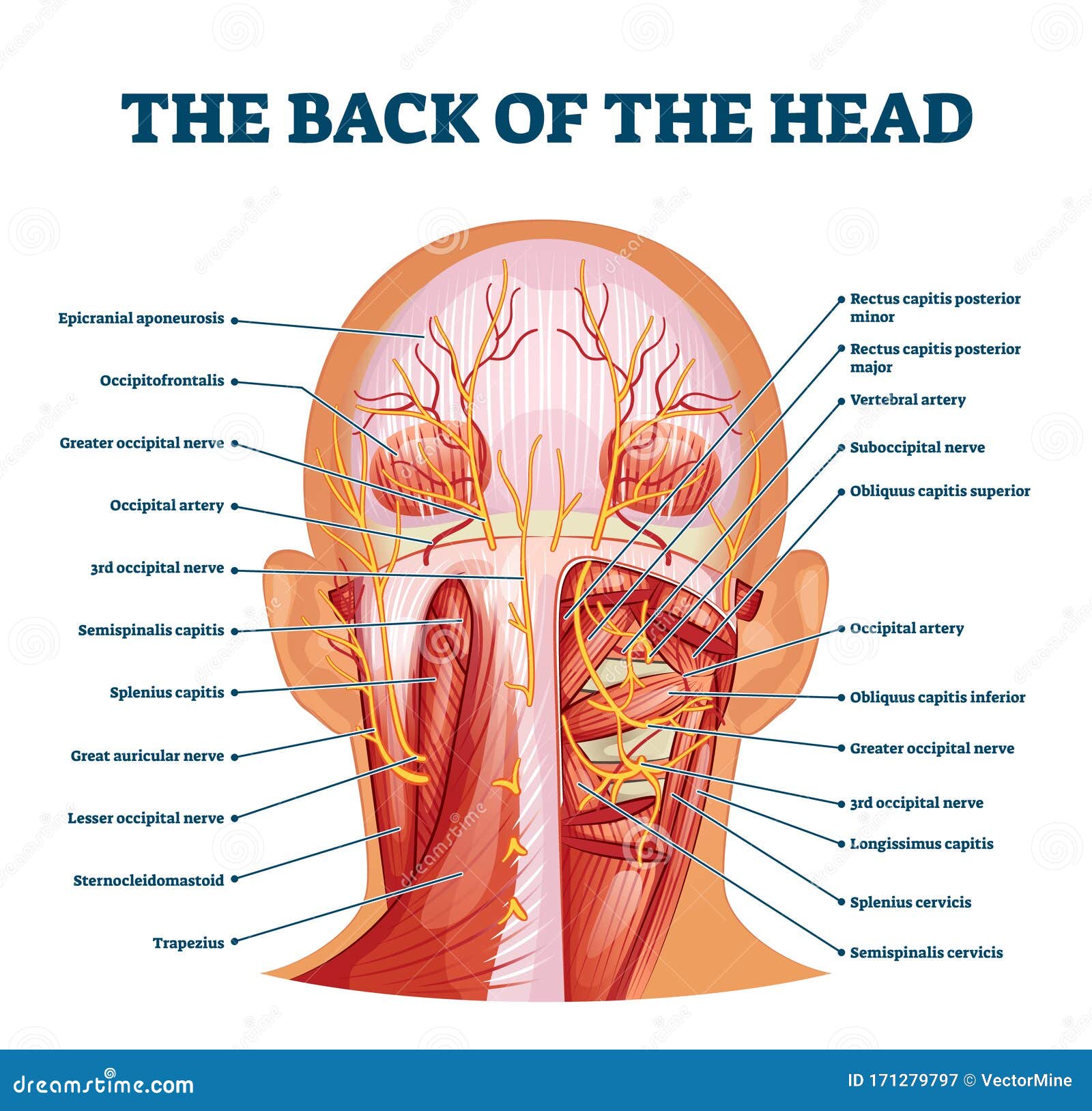
Back Of The Head Muscle Structure And Nerve System Diagram Stock Vector Illustration Of Labeled Muscle 171279797
Muscles Of The Head Neck And Back Illustrations Radiology Case Radiopaedia Org
Ppt Head Neck Shoulder Back Muscles Powerpoint Presentation Id 9122180

The Human Muscle System Neck Muscle Anatomy Muscles Of The Neck Muscle Anatomy

Dentistry And Medicine Head And Neck Anatomy Muscles Blood Supply Diagrams Free Download

Posterior Triangle Of The Neck Head And Neck Anatomy Human Body Muscle Neck Muscle Text Hand Human Png Pngwing
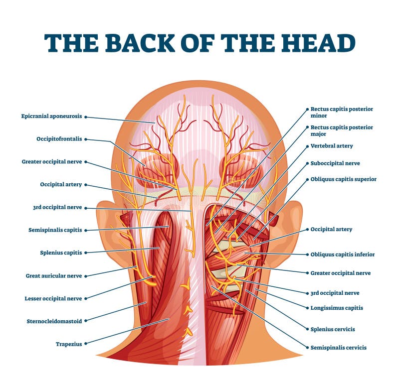
Back Of The Head Muscle Structure And Nerve System Diagram Stock Vector Illustration Of Labeled Muscle 171279797
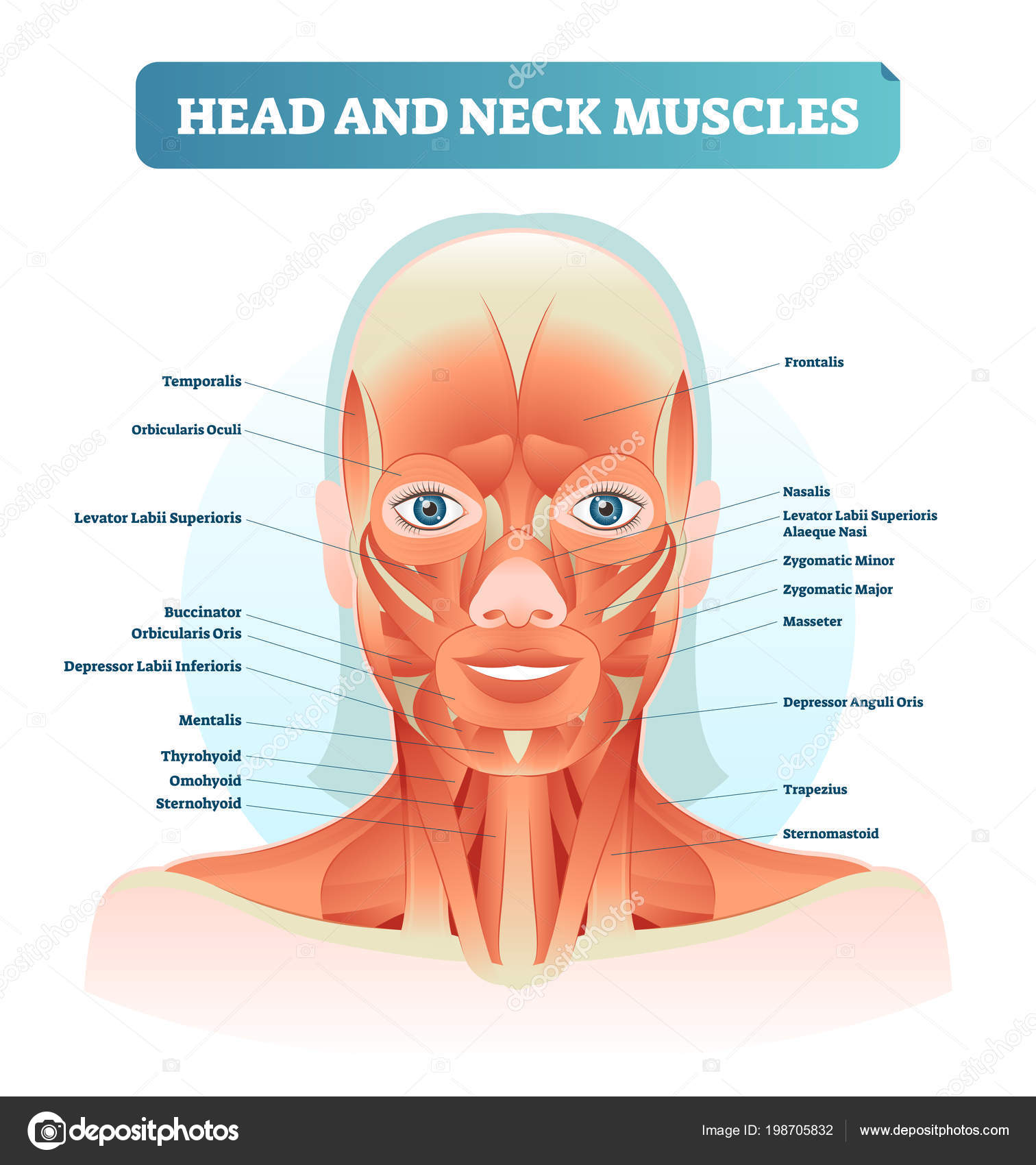
Head And Neck Muscles Labeled Anatomical Diagram Facial Vector Illustration With Female Face Health Care Educational Information Poster Stock Vector Image By C Vectormine 198705832
0 Response to "45 diagram of neck muscles"
Post a Comment