44 drag the labels onto the diagram to identify the types of connective tissue proper.
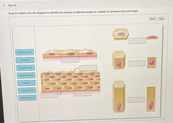
Part A Drag the labels onto the diagram to identify the types of epithelia. ANSWER: Correct Art-labeling Activity: Types of Connective Tissue Proper Learning Goal: To learn the types of connective tissue proper. Label the types of connective tissue proper. Determine which connective tissue type each image below represents. Then click and drag the labels matching them up with the correct tissue type. ... Identify each tissue type pictured. Then click and drag each label to the tissue type it describes. Click and drag the words or phrases into the appropriate sentences to describe glands.
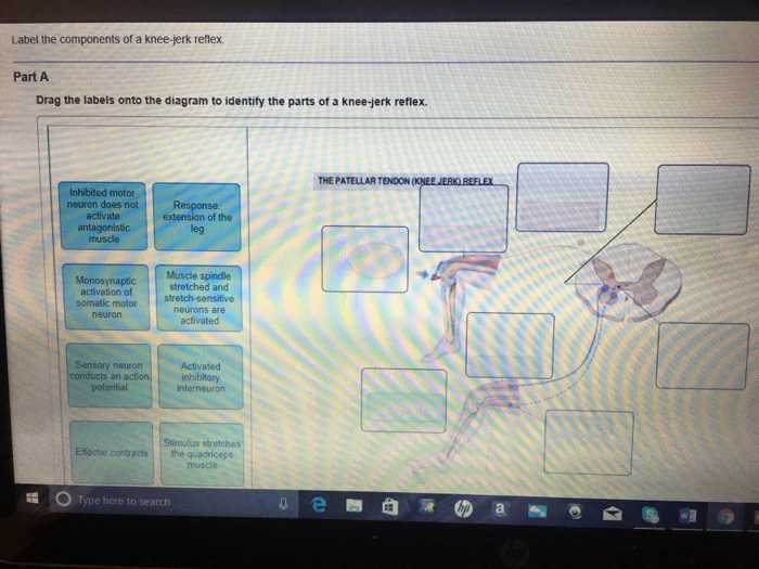
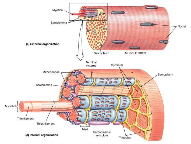
These tissues include the skeletal muscle fibers blood vessels nerve fibers and connective tissue. Drag the labels onto the diagram to identify structural features associated with skeletal muscle. Drag the labels onto the diagram to identify the blood types that correspond to specific blood typing test results.

Drag the labels onto the diagram to identify the types of connective tissue proper.
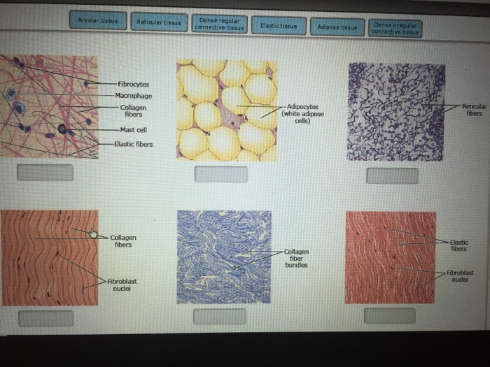
Drag the labels onto the diagram to identify the types of connective tissue proper. areolar tissue, adipose tissue, reticular tissue, dense regular connective tissue, dense irregular connective tissue, elastic tissue. Myosatellite cells are found in association with _____ muscle. The word tissue derives from the Old French word meaning "to weave," reflecting the fact that the different tissues are woven together to form the "fabric" of the human body. The four basic types of tissue are epithelial tissue, connective tissue, muscle tissue, and nervous tissue. If a single, broad functional term were assigned to ... Connective tissue is a term used to describe the tissue of mesodermal origin that that forms a matrix beneath the epithelial layer and is a connecting or supporting framework for most of the organs of the body. This lab will focus on the so-called connective tissue proper and cartilage; the next lab will focus on bone.
Drag the labels onto the diagram to identify the types of connective tissue proper.. Identify the muscle tissue type described by choosing the correct response(s) from the key choices. Enter the appropriate term(s) or letter(s) of the key choice in the answer blank. Key Choices A. Cardiac B. Smooth C. Skeletal Banded appearance Longitudinally and circularly arranged layers Dense connective tissue packaging Identify the tissue type and a location where it is found. Loose Areolar Connective Tissue •Papillary layer of dermis • Hypodermis •Around organs • Basement membrane of mucous membranes •Surrounding blood vessels Blood Vessel. Identify the tissue type and its function. The dermis is subdivided into a thin layer of loose connective tissue atop a thicker dense connective tissue, which contains most of the glands and other structures, including hair follicles. Match the layers and structures in the diagram to the proper terms. Tough Topics: The Structure of Skin-- Layers and Tissues The integument is absolutely ... Each skeletal muscle has three layers of connective tissue that enclose it, provide structure to the muscle, and compartmentalize the muscle fibers within the muscle (Figure 10.2.1). Each muscle is wrapped in a sheath of dense, irregular connective tissue called the epimysium , which allows a muscle to contract and move powerfully while ...
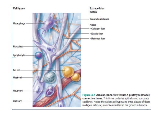
A basic microscope has a single convex lens such as those found in a magnifying glass which you can use to visualize the finest prints Drag the labels onto the diagram to identify the types of bone cells osteogenic cell osteocyte osteoblast osteoclast Drag the labels to the appropriate location in the figure Lacunae Lamellae Osteon Identify the ... Mastering ap exam 1. Drag the labels onto the diagram to identify the stem cells and stages of white blood cell and pl. Label the components of a model cell. Dna polymerase begins synthesizing the lagging strand by adding nucleotides to a short segment of rna. Na is entering the cell. (c) Connective tissue proper: loose connective tissue, reticular Description: Network of reticular fibers in a typical loose ground substance; reticular cells lie on the network. Function: Fibers form a soft internal skeleton (stroma) that supports other cell types including white blood cells, mast cells, and macrophages. Drag the labels onto the diagram to identify the types of connective tissue proper. look at pic When a molecule is _______ it loses electrons, and when a molecule is ______ it gains electrons.
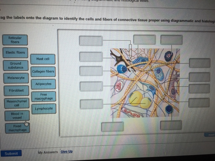
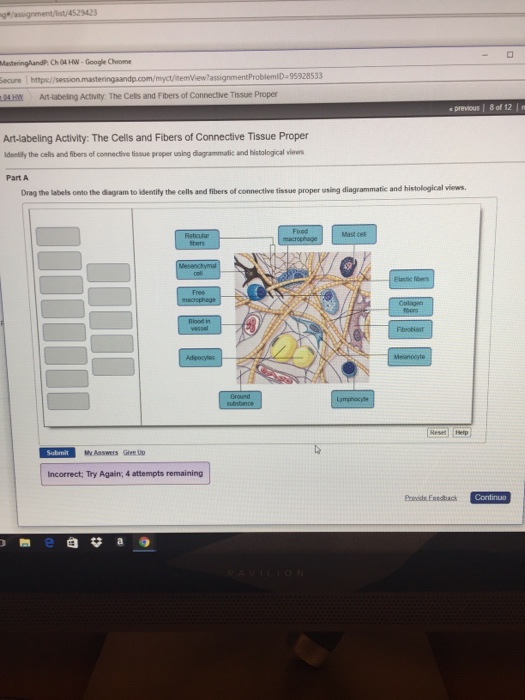
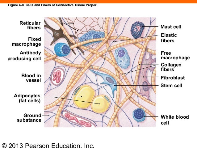
100% (5 ratings) Answer There types of tissues included in connective tissue proper are : Loose (areolar) con …. View the full answer. Transcribed image text: Drag the labels onto the diagram to identify the cells and fibers of connective tissue proper using diagrammatic and histological views. Previous question Next question. Drag the labels onto the diagram to identify the types of connective tissue proper. (week three assignment one) #7. Drag the labels onto the four types of tissue membranes. ... Drag the labels onto the diagram to identify the effects of isotonic, hypotonic, and hypertonic solutions on red blood cells. The major types of connective tissue are connective tissue proper, supportive tissue, and fluid tissue. Loose connective tissue proper includes adipose tissue, areolar tissue, and reticular tissue. These serve to hold organs and other tissues in place and, in the case of adipose tissue, isolate and store energy reserves. Drag the correct description under each cell structure to identify the role it plays in the cell. Part a animal cell structure drag the labels onto the diagram to identify the structures of an animal cell. Drag the labels onto the diagram to identify the parts of the cell. Labels can be used once more than once or not at all.
31 Drag The Labels Onto The Diagram To Identify The Types ...
Transcribed image text: Drag the labels onto the diagram to identify the structural classification of exocrine glands. Res Compound Dubular Simple coiled Oland Compound Alveolar (acinar) Simple tubular Simple branched hiver Simple branched Simple alveo (honan Compound . Previous question Next question. COMPANY.
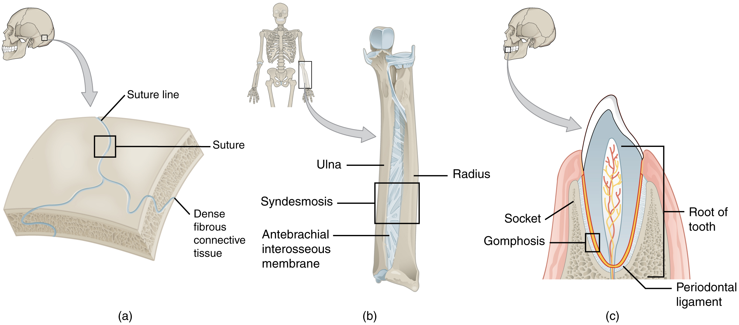
Fibrous Joints · Anatomy and Physiology
View Homework Help - chapter 4.pdf from AAS a&p 198 at University of New Hampshire. 12/24/13 chapter 4 chapter 4 Due: 9:00am on Thursday, September 26, 2013 You will receive no credit for items you
Solved: Drag The Labels Onto The Diagram To Identify The D ...
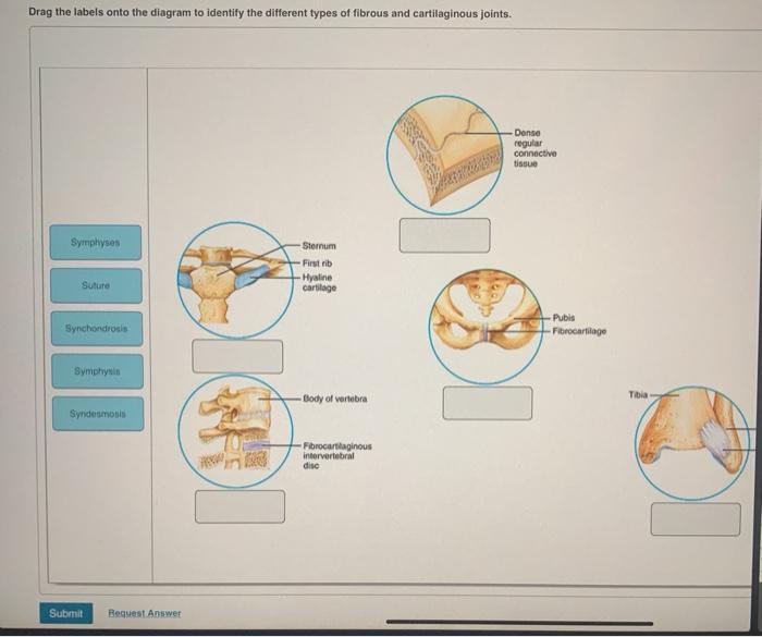
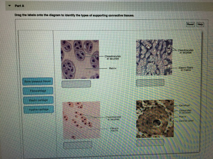
Drag the labels onto the diagram to identify the types of supporting connective tissues. look at pic A marked loss in strength and elasticity of connective tissue characterizes Marfan's syndrome.

Solved: Drag The Labels Onto The Diagram To Label The Step ...
Sounds a little weird, but your bones and your blood are just types of connective tissue! 02:48 So, despite the name, your connective tissues do way more than just connect your muscles to your bones.

Anatomy And Physiology Archive | September 02, 2018 ...
Anatomy and Physiology questions and answers. Drag the labels onto the diagram to identify structures of the urinary bladder. Re Basal lamina Connective tissue Stretched bladder Epithelium (stretched) Relaxed bladder Epithelium (relaxed) Question: Drag the labels onto the diagram to identify structures of the urinary bladder.

Drag The Labels To Identify The Structures Of A Long Bone ...
Connective tissue is a term used to describe the tissue of mesodermal origin that that forms a matrix beneath the epithelial layer and is a connecting or supporting framework for most of the organs of the body. This lab will focus on the so-called connective tissue proper and cartilage; the next lab will focus on bone.
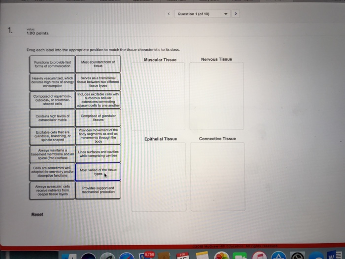
Solved: Question 1 (of 10) 100 Points Drag Each Label Into ...
The word tissue derives from the Old French word meaning "to weave," reflecting the fact that the different tissues are woven together to form the "fabric" of the human body. The four basic types of tissue are epithelial tissue, connective tissue, muscle tissue, and nervous tissue. If a single, broad functional term were assigned to ...
Solved: EwassignmentProblemID 76141051 1 A&P I Fall 2016 S ...
Drag the labels onto the diagram to identify the types of connective tissue proper. areolar tissue, adipose tissue, reticular tissue, dense regular connective tissue, dense irregular connective tissue, elastic tissue. Myosatellite cells are found in association with _____ muscle.

Drag The Labels Onto The Diagram To Identify Structural ...
Drag The Labels Onto The Diagram To Identify Structural ...
Anatomy And Physiology Archive | September 18, 2017 ...

Drag The Labels Onto The Diagram To Identify The ...

280 best images about Central Nervous System on Pinterest ...

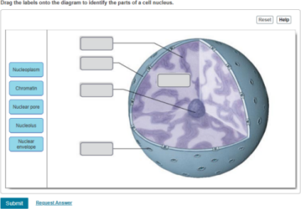
Drag The Labels Onto The Diagram To Identify The Parts Of ...
163 ch 04_lecture_presentation

Drag The Labels Onto The Diagram To Identify Structural ...
Drag The Labels Onto The Diagram To Identify The ...
Drag The Labels Onto The Diagram To Identify The Types Of ...

Drag The Labels Onto The Diagram To Identify The ...

Drag The Labels To Identify The Structures Of A Long Bone ...
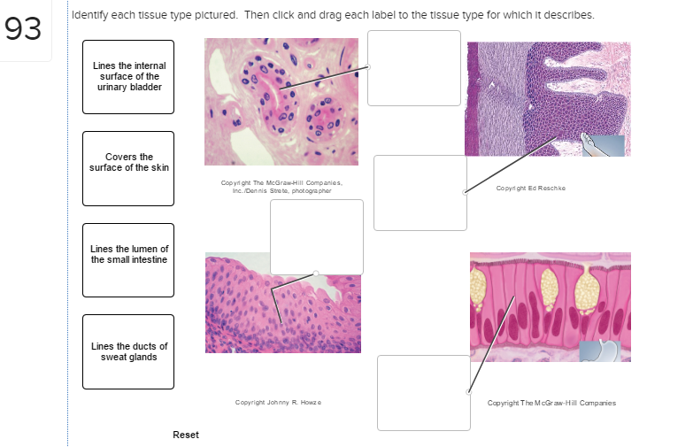
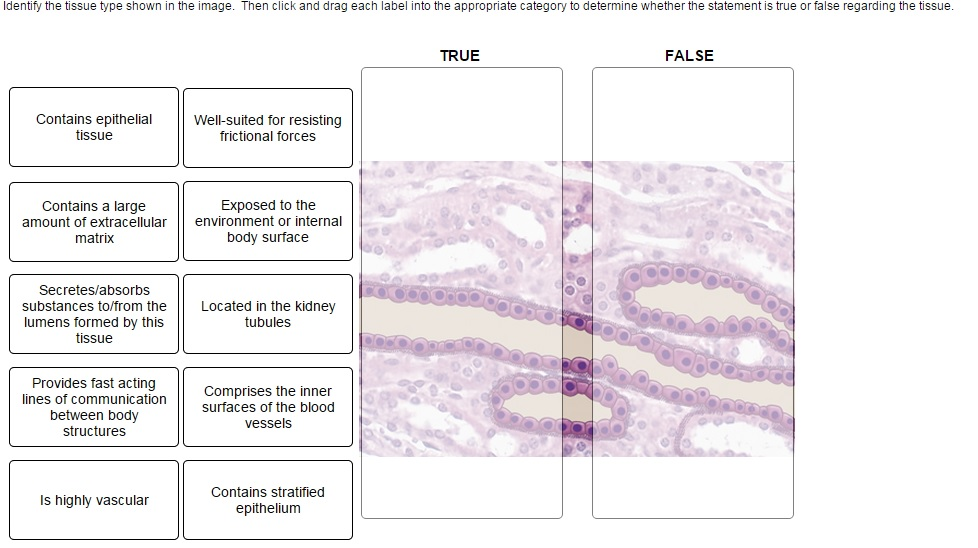
Solved: Identify Each Tissue Type Pictured. Then Click And ...
33 Drag The Labels Onto The Diagram To Identify The Types ...

Tissues of the Body Flashcards | Quizlet
Anatomy & physiology for pg dentistry 01
Solved: -Aibrocytes Reticular Fbers Bundles Part A Drag Th ...

Drag The Labels Onto The Diagram To Identify The Parts Of ...

31 Drag The Labels Onto The Diagram To Identify The Types ...

Drag The Labels Onto The Diagram To Identify The ...
Part A Drag the labels onto the diagram to identify ...
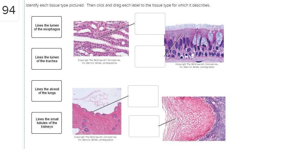
Solved: Identify Each Tissue Type Pictured. Then Click And ...

84 best Connective tissues images on Pinterest | Human ...
Drag The Labels Onto The Diagram To Identify The Parts Of ...
Connective Tissue Diseases : Facts, types, symptoms ...
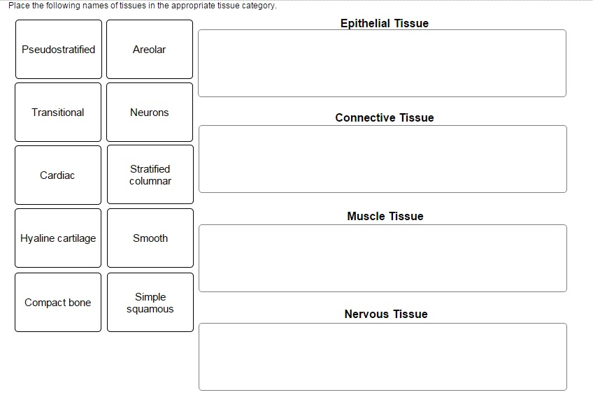
Solved: Place The Following Names Of Tissues In The Approp ...

Which Layer Is Composed Primarily Of Dense Irregular ...
Solved: Drag Each Label Into The Appropriate Position To M ...

30 Drag The Labels Onto The Diagram To Identify The Types ...
30 Drag The Labels Onto The Diagram To Identify Structural ...

EDS on a cellular level
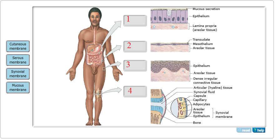
Solved: Mucous I Membrane ? Cutaneous Membrane Serous Memb ...

Drag the labels onto the diagram to identify the tissues ...
Solved: EwassignmentProblemID 76141051 1 A&P I Fall 2016 S ...

Drag The Labels Onto The Diagram To Identify Structural ...

Solved: Drag The Labels Onto The Diagram To Identify The C ...
0 Response to "44 drag the labels onto the diagram to identify the types of connective tissue proper."
Post a Comment