44 elodea leaf cell diagram
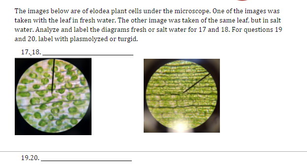
Using a compound light microscope, a student observes the reaction of an Elodea leaf’s cell to a 10% sucrose solution. After a few minutes, she notices that the cell membrane pulls away from the cell wall. She sketches her observations. ... In the diagram, cells with an internal salt concentration of 5% are placed in two solutions. Solution A ...
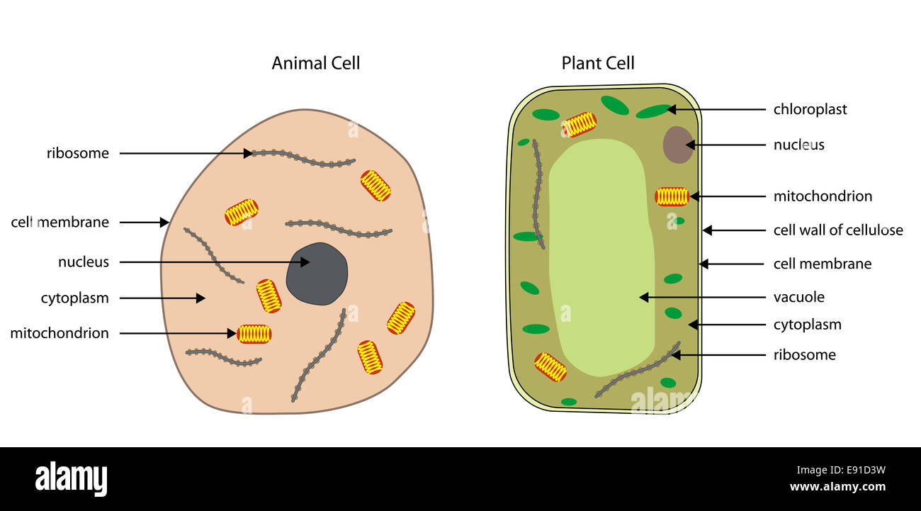
Animal cells vs. Plant cells – Key similarities Animal cells and plant cells are eukaryotic cells. Both animal and plant cells are classified as “Eukaryotic cells,” meaning they possess a “true nucleus.”Compared to “Prokaryotic cells,” such as bacteria or archaea, eukaryotic cells’ DNA is enclosed in a membrane-bound nucleus.These membranes are similar to the cell membrane ...
Gizmo Warm-up In the Cell Types Gizmo, you will use a light microscope to compare and contrast different samples. On the LANDSCAPE tab, click on the Elodea leaf. (Turn on Show all samples if you can’t find it.) Switch to the MICROSCOPE tab to observe the sample as it would appear under the microscope.

Elodea leaf cell diagram
Elodea Leaf Cell Diagram. 1 b Give the. A List A gives the names of three parts of the cell. Nov 22 2017 - This Pin was discovered by Sarah Shamra. In this article we will discuss about the structure of a leaf with the help of a diagram. Plant leaves help to sustain life on earth as they generate food for both plant and animal life.
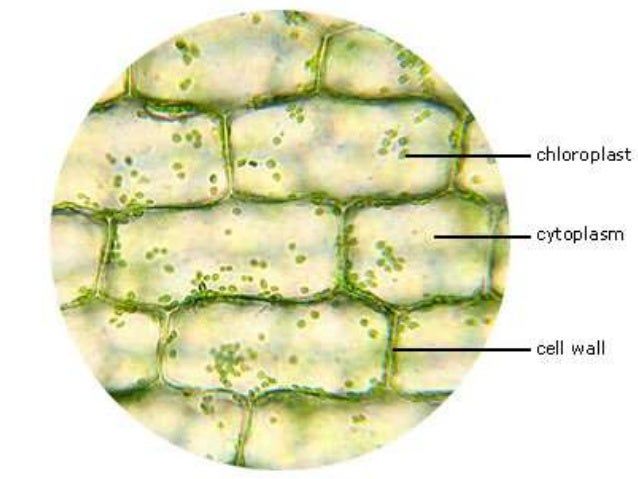
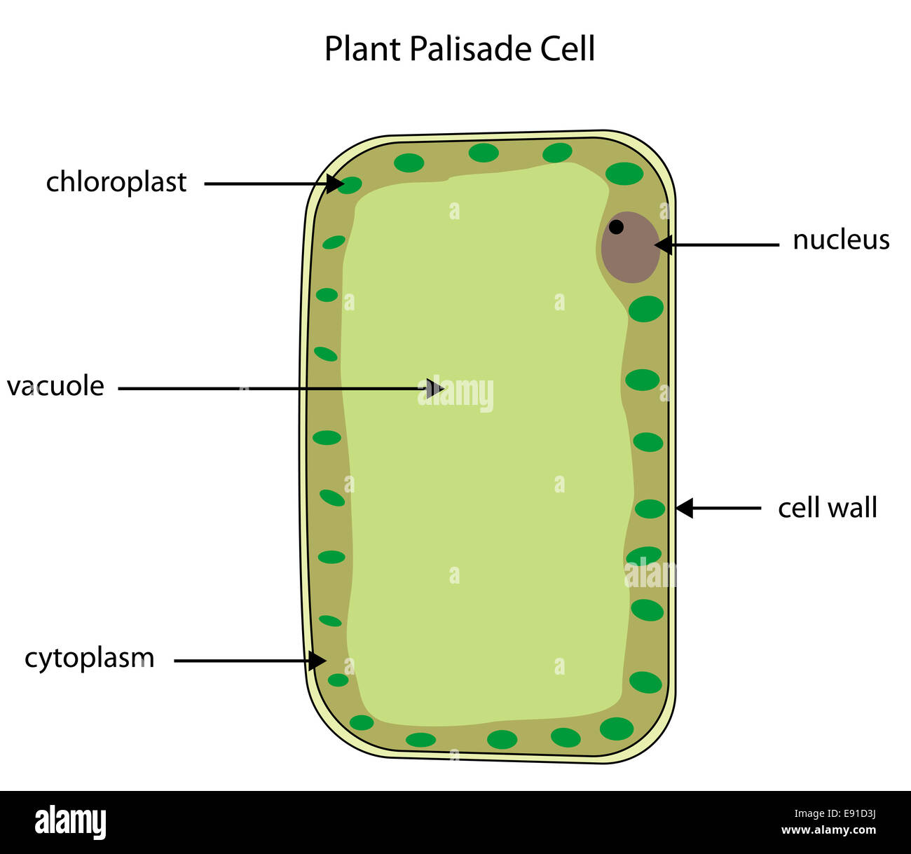
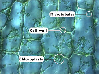
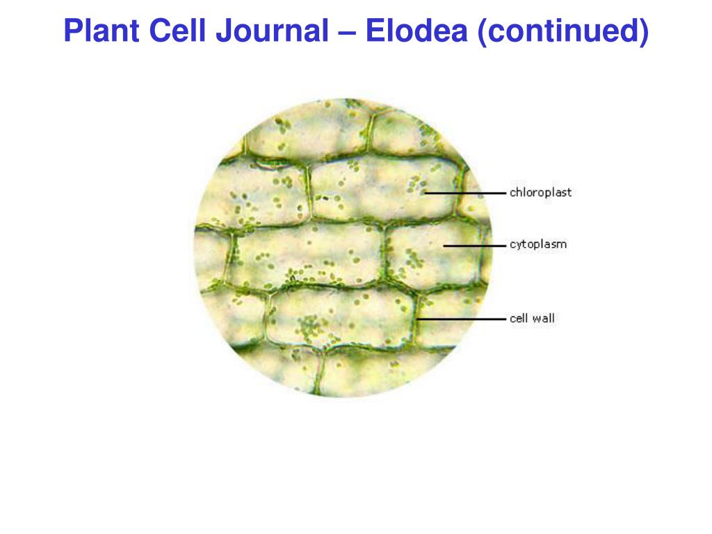
What is a Elodea cell? This Elodea leaf cell exemplifies a typical plant cell. It has a nucleus, and a stiff cell wall which gives the cell its box-like shape. The numerous green chloroplasts allow the cell to make its own food (by photosynthesis). The central vacuole takes up most of the volume of the cell.
Plasmolysis In Plant Cell Diagram. The cells with only cell membranes, such as animal cells, eventually swell and burst. This indicates the phenomenon of deplasmolysis. A bacteria diagram clearly enables us to profit extra about this unmarried cell organisms that have neither membrane-bounded nucleolus or organelles like mitochondria and chloroplasts. They are obviously a trigger […]
Elodea leaf cell diagram.
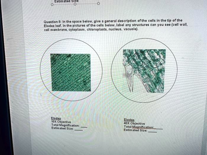
To create a wet mount of an Elodea leaf, this general procedure was followed. Put a drop of water on the center of a slide. Cut an extremely small piece of an Elodea leaf and place it on the water. Place a coverslip onto the slide. Observe the plant cell slide under the virtual microscope by following this procedure.
Elodea Leaf Cell Diagram. In a plant cell the cell wall is made up of cellulose hemicellulose and proteins while in a fungal cell it is composed of chitin. Centrioles help move chromosomes during cell division. Animal Cell Labeled Science At Co Op Animal Cell Plant Cell.
Chloroplast division occurs every cell cycle right before the separation of two daughter cells (cytokinesis). Cut a small piece of Elodea leaves and put on the slide. How many chloroplasts can be found in one cell? A typical animal cell is 10-20 μm in diameter, which is about one … When thin sections of a chloroplast are examined under the electron microscope, several features are ...
Turgor pressure is the force within the cell that pushes the plasma membrane against the cell wall.. It is also called hydrostatic pressure, and is defined as the pressure in a fluid measured at a certain point within itself when at equilibrium. Generally, turgor pressure is caused by the osmotic flow of water and occurs in plants, fungi, and bacteria. ...
The plant cell wall is a barrier to virus exit from and entry into cells. Thus, successful infection requires that plant viruses encode movement proteins to transport the viral genome locally cell-to-cell within a leaf and on into the vascular system, through which the virus will systematically invade the plant.
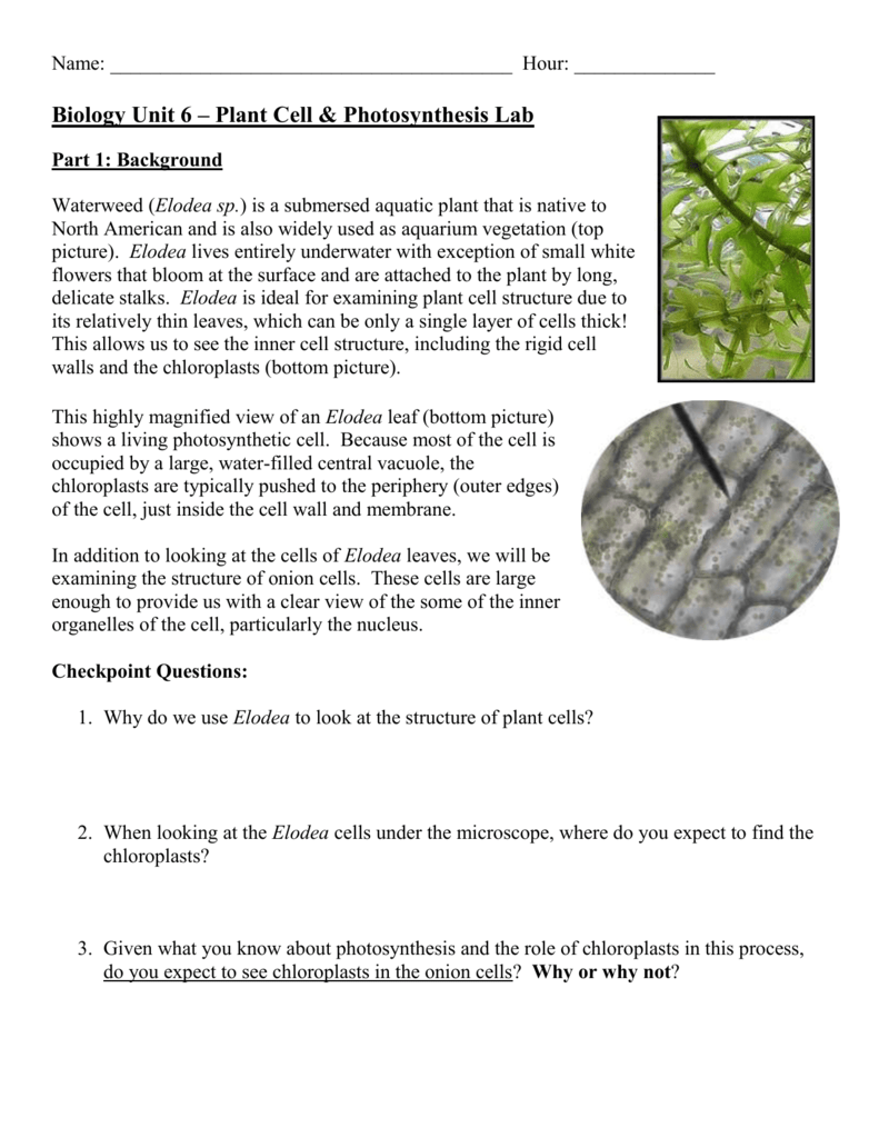
The Elodea leaf is composed of two layers of cells. It is similar to a disc with a central depression. The structure labeled G give rise to spindle fibers and exclusively seen in animal cell. We hope this picture Plant Cell Diagram Label can help you study and research.
Video description:-Video describes 3 minutes of continuous Elodea plant cell observations. Put an Elodea leaf in a drop of water on a slid. Observe under the microscope and draw just ONE good cell. 2014 Apr 13 - Elodea leaf plant cell under the microscope. They are connected by stroma lamellae.. Slice of onion.
A. diagram 1 could represent carbon dioxide exiting a leaf cell during photosynthesis. B. diagram 2 could show facilitated diffusion of urea out of an active muscle cell. C. diagram 3 could represent pumping potassium ions into a cell. D. diagram 4 could represent oxygen diffusing into a blood cell. Question 3
Elodea leaf: Elodea leaves are two cell layers thick. Either the nucleus of an Elodea cell or cytoplasmic streaming of the chloroplasts in an Elodea cell. Magnification x1000 to 1500 Can be used to see organisms up to 1 millionth of meter but resolution is not as high and clear. Place an Elodea leaf in the drop of water, place a coverslip on top.
It is also often used in biology labs on cell structure because it forms nice, large cells that are easily observed under the microscope. In this virtual lab, you will view cells from your cheek animal cells and cells from onion, and from elodea, which is a water plant. Cells are joined together to make up the leaf tissue.
Why are elodea cells rectangular? This Elodea leaf cell exemplifies a typical plant cell. It has a nucleus, and a stiff cell wall which gives the cell its box-like shape. The numerous green chloroplasts allow the cell to make its own food (by photosynthesis). The central vacuole takes up most of the volume of the cell.
Onion cells are brick-like in shape while human cheek cells are rounded. Human cheek cells do not have a cell wall or a large vacuole. Which of the following is a main difference in cell structure between an elodea plant cell and a human cheek animal cell? 1 Answer. The elodea cell will have a central vacuole in addition to chloroplasts.
The raw materials of photosynthesis, water and carbon dioxide, enter the cells of the leaf, and the products of photosynthesis, sugar and oxygen, leave the leaf. Cross section of a leaf, showing the anatomical features important to the study of photosynthesis: stoma, guard cell, mesophyll cells, and vein.
Contractile Vacuole 2 Peace Symbol Biology Symbols. Animal Vs Plant Cells Nice Unlabeled Diagrams Exploring Nature Plant Cell Diagram Cell Diagram Plant Cell. Elodea Water Plant Under Microscope Cell Walls And Chloroplasts Are Clearly Vis Sponsored Microscope Cell Walls E Water Plants Cell Wall Plant Cell. In cell, diagram, elodea.
This Elodea leaf cell exemplifies a typical plant cell. It has a nucleus, and a stiff cell wall which gives the cell its box-like shape. The numerous green chloroplasts allow the cell to make its own food (by photosynthesis). Like animal cells, the cytoplasm of this plant cell is bordered by a cell membrane. Is an Elodea leaf cell an organism?
A cell is a very tiny structure which exists in living bodies. These cells combine to form tissues, and later the tissues combine to form different the basic structure of the animal and plant cell is quite similar as both of them are eukaryotic cells. Golgi bodies are frequently seen here under the microscope. Source: image.shutterstock.com
It's quite difficult for me to learn the way Osmosis work. Osmosis and Diffusion. 4K answer views. Osmosis - Elodea Lab 10. Predict the end result of placing a red blood cell or Elodea plant cell into these situations. #4112. of the Cell Sap 4. The weakest being 0%, and the highest being 5%. Some organisms have cell vacuoles also.
A Fishbone Diagram Example Heres a completed version to help show you what to expect. Perhaps for example using the 4M method. Here is an example of a completed fishbone diagram showing information entered for each of the four categories agreed upon by this team. Draw a straight line from the head leading to the left.
6. The diagram represents part of a life process in a leaf chloroplast. If the process illustrated in the diagram is interrupted by a chemical at point X, there would be an immediate effect on the release of A. chlorophyll B. nitrogen C. carbon dioxide D. oxygen 7. The equation below represents a summary of a biological process.
Jul 19, 2021 · Basis Prokaryotic Cell Eukaryotic Cell; 1. Examples: Cells of bacteria, archaea or archaebacteria, and cyanobacteria or blue-green algae: Cells of plants, animals, fungi, algae, and protozoa ()2. Type: Unicellular: Unicellular or multicellular
Cells are joined together If you look closely, you can see, the cells chloroplasts moving throughout the cell. Download the royalty-free video "movement of chloroplasts with chlorophyll in leaf cells of elodea under microscope 4K " created by klickit24 at. Today you will be working with the aquatic plant Elodea, also known as anacharis.
Osmosis Elodea Lab. Label and draw the elodea leaf under high power: Figure 4:. Cell Structure. It follows that the specimen must be carefully prepared, or 'mounted' on a slide. Simply mount a whole leaf in water under a cover slip. You should see the chloroplasts moving in a circular pattern around the cell.
About Under Elodea The Microscope . The Magnification of 400x is the best level for best view leaf cell structure. Procedure A. of Elodea under the light for 15-20 minutes. Look at the leaf under the microscope (Figure 4. Elodea, a common aquarium plant, has very thin leaves and is a good specimen for studying the cells of green plants.
Elodea Leaf Cell Diagram. Draw A Labelled Diagram Of The External Structure Leaf Brainly In. It is the stalk that connects a leaf to the stem of the plant it is made of complex conducting tissues called vascular tissues. This is a quiz called leaf structure and was created by member synyster sik. KS3 Y7 History Medieval Early Modern History.
BIOLOGY 101 Cell Types Gizmos 2018 Activity A: Observing cells Get the Gizmo ready: On the LANDSCAPE tab, click on the woman's right arm to choose the Human skin sample. Select the MICROSCOPE tab. Introduction: Complex organisms are made up of smaller units, called cells. Most cells are too small to be seen by the naked eye.
Elodea Leaf Cell Diagram. Notice distinct nucleus, cytoplasm and cell membrane. Tinción: H-E, 100X. The scale bar has a width of 20 micrometers. The Elodea leaf is composed of two layers of cells. The reason that an elodea contains a chloroplast is due to photosynthesis.
Elodea cells have a cell wall, a central vacuole, and chloroplasts, but cheek cells do not have these structures. The structure in a cheek cell appearing darker by absorbing most of the stain is the _____.
The boxes and that you see in the elodea are cells. Cells are joined together If you look closely, you can see, the cells chloroplasts moving throughout the cell. Download the royalty-free video "movement of chloroplasts with chlorophyll in leaf cells of elodea under microscope 4K " created by klickit24 at.
The diagram below represents the change that occurred after a fluid was added to a wet mount of some ELODEA leaf cells. In the diagram below the two blue strands represent. 2 in the diagram below the green unit represents. Part c in the diagram below the two blue strands represent. Lec 6 mastering biology. Activity 17b the terminator.
Procedure: Prepare a wet mount of one leaf from the water plant Elodea using the water in which it is kept. Observe the cells under normal conditions, and make a sketch of what you see. While observing the leaf under the microscope, wick a solution of 6% NaCl (sodium chloride) across the slide. Sketch your observations.
November 16, 2021. Elodea Leaf Cell Under Microscope Plant Cell Cells Worksheet Lab Activities. Cell Transport Lab Osmosis And Diffusion Cell Transport Cell Osmosis. Plant Cells Vs Animal Cells With Diagrams Animal Cell Plant Cell Diagram Plant Cell. Plant Cell Electron Microscope Worksheet Cell Diagram Plant Cell Plant Cell Diagram.
The Elodea leaf consists of two layers of cells. Solely one layer ofcells is in focus when utilizing the excessive energy (40x) goal. Click on to see full reply. Subsequently, one can also ask, how large is an elodea cell? The plasma membrane is just too skinny to see at this magnification. Within the printed picture the scholars work with, the ...
The boxes and that you see in the elodea are cells. Cells are joined together If you look closely, you can see, the cells chloroplasts moving throughout the cell. Download the royalty-free video "movement of chloroplasts with chlorophyll in leaf cells of elodea under microscope 4K " created by klickit24 at.
/plant-cell-elodea--isotonic-solution-shows-cells--chloroplasts-250x-at-35mm-139802547-5a956de86bf069003717851a.jpg)












0 Response to "44 elodea leaf cell diagram"
Post a Comment