43 simple columnar epithelium labeled diagram
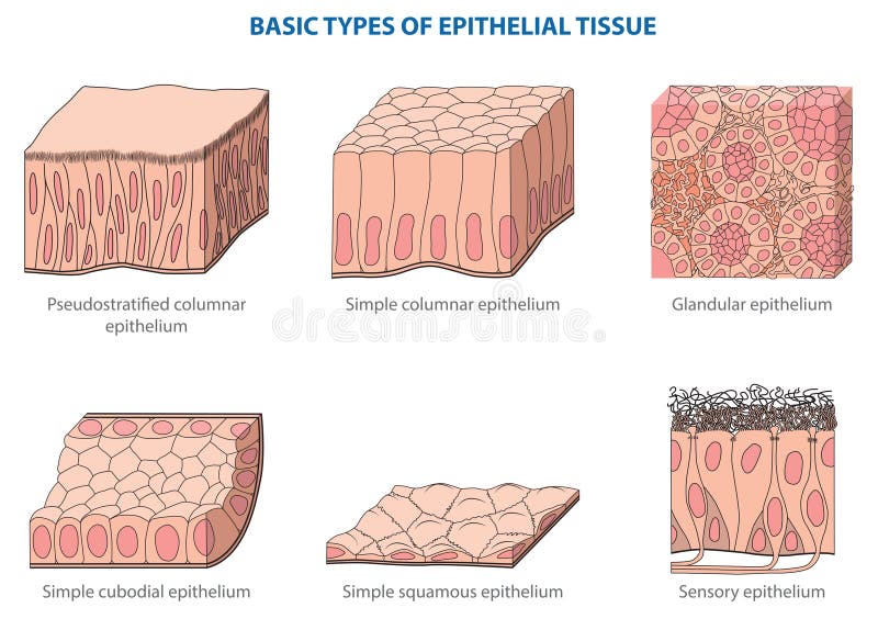
Lab 2: Microscopy and the Study of Tissues - UW-La Crosse Simple epithelium is composed of a single layer of cells while stratified epithelium contains several layers. Epithelial sells can be flat (squamous = "scale-like"), cube-shaped (cuboidal) or tall (columnar). So, to correctly identify the type of tissue requires three words (e.g., simple columnar epithelium, stratified, squamous epithelium, etc. PDF Practice Quiz Tissues - Portland Community College Nonciliated Simple Columnar Epithelium •Absorption •Secretion of mucus, enzymes, and other substances. Identify the tissue type and a location where it is found. Nonciliated Simple Columnar Epithelium •Lines most of the digestive tract •Excretory ducts of some glands.
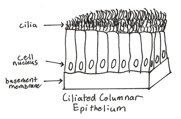
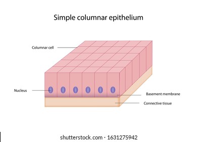
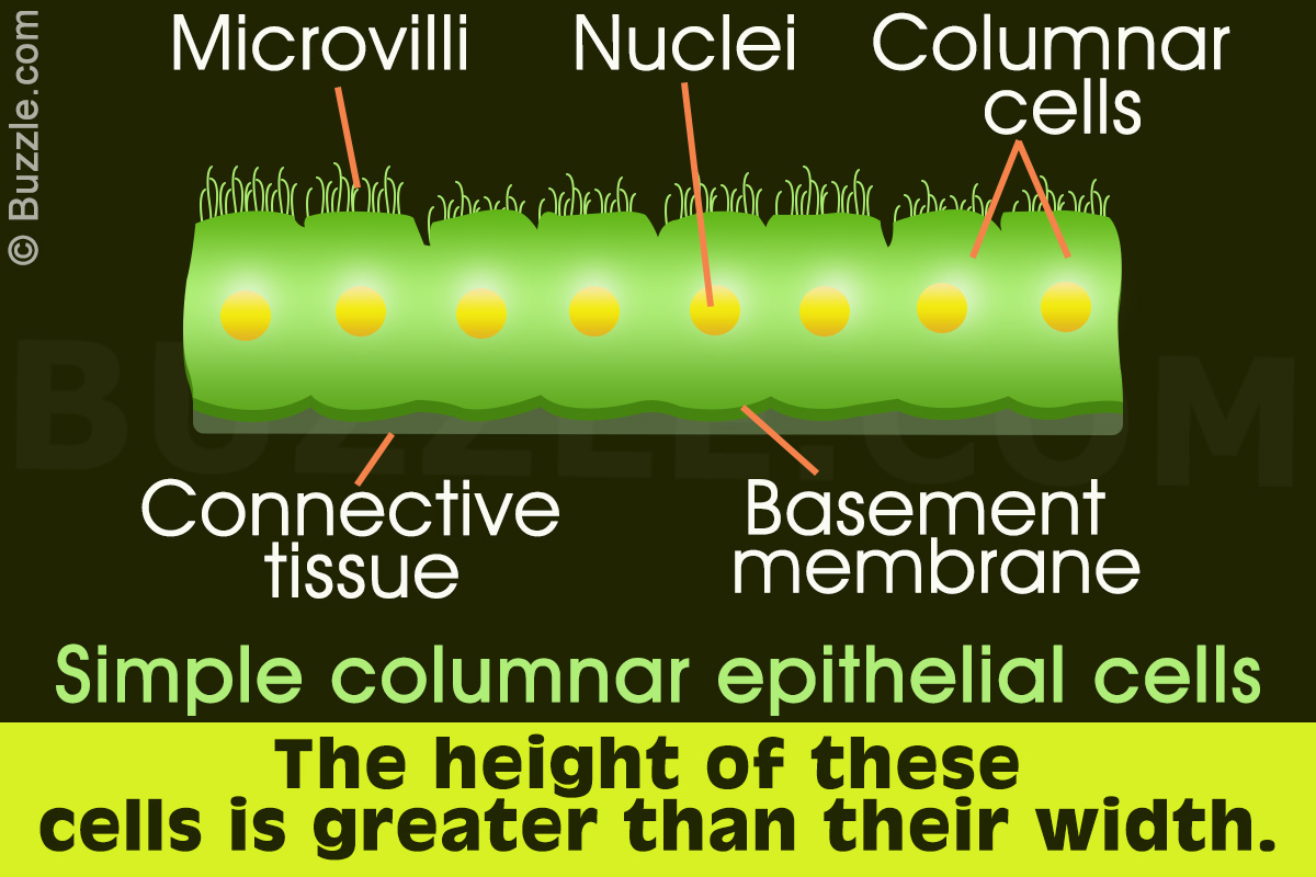
4.2 Epithelial Tissue - Anatomy & Physiology Columnar epithelia, which form the lining of the digestive tract, can be either simple or stratified. The cells are long and narrow. The nucleus is elongated and located on the basal side of the cell. Ciliated columnar epithelium is composed of simple columnar epithelial cells that display cilia on their apical surfaces.
Simple columnar epithelium labeled diagram
Simple Columnar Epithelium Labeled Diagram Simple Columnar Epithelium Labeled Diagram Ciliated columnar epithelium is composed of simple columnar epithelial cells with cilia on their apical This illustration shows a diagram of a goblet cell. These labelled diagrams should closely follow the current Science (simple squamous epithelium). ORIGIN: columnar epithelium with goblet cells. TISSUE . PDF Marieb HA8 chapter 4 - Pearson cells. This epithelium forms the secretory cells of many glands, the walls of the smallest ducts of glands, and the walls of many tubules in the kidney. Its functions are the same as those of simple columnar epithelium. Simple Columnar Epithelium (Figure 4.3c) Simple columnar epithelium is a single layer of tall cells aligned like soldiers in a ... Epithelial Tissue - Anatomy and Physiology Like the cuboidal epithelia, this epithelium is active in the absorption and secretion of molecules. Simple columnar epithelium forms the lining of some sections of the digestive system and parts of the female reproductive tract. Ciliated columnar epithelium is composed of simple columnar epithelial cells with cilia on their apical surfaces.
Simple columnar epithelium labeled diagram. Simple Squamous Epithelium Function: Passage of materials ... Simple Squamous Epithelium Function: Passage of materials by diffusion and filtration Location: Air sacs of lungs ... Location: Glandular tissue, kidney tubules Simple Columnar Epithelium Function: Absorption Location: Lining of the digestive tract . Pseudostratified Columnar Epithelium Function: Secretion, particularly mucus Location: Lining ... View 11 Simple Columnar Epithelium Location - ravinetinesz Simple Columnar Epithelium Location are a subject that is being searched for and appreciated by netizens these days. You can Get the Simple Columnar Epithelium Location here. Get all royalty-free pix. We Have got 12 pix about Simple Columnar Epithelium Location images, photos, pictures, backgrounds, and more. In such page, we additionally have ... Simple Columnar Epithelium: A Labeled Diagram and ... Simple Columnar Epithelium: A Labeled Diagram and Functions Epithelium is a tissue that lines the internal surface of the body, as well as the internal organs. Simple epithelium is one of the types of epithelium that is divided into simple columnar epithelium, simple squamous epithelium, and simple cuboidal epithelium. Jejunum Histology Slide with Labeled Diagram and ... The lining epithelium of the tunica mucosa is a simple columnar epithelium with goblet cells. You may also find other different cells in the mucosa of the small intestine like penath cells, enteroendocrine cells, and others. The number of the plica circularis and villi mary varies in the different parts of an animal's small intestine.
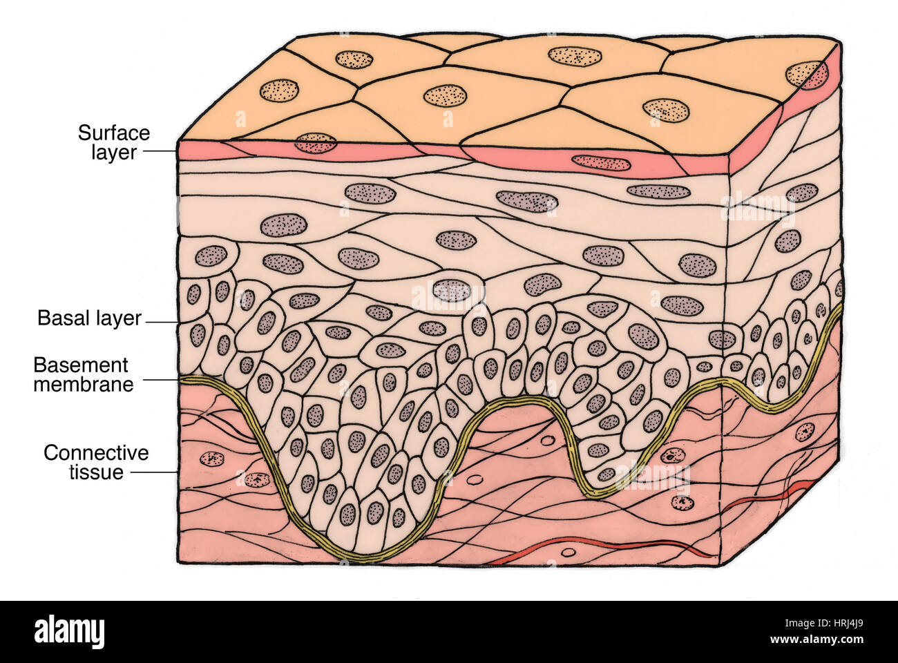
Simple epithelium: Location, function, structure | Kenhub Simple epithelium can be divided into 4 major classes, depending on the shapes of constituent cells. The cells found in this epithelium type are flat and thin, making simple squamous epithelium ideal for lining areas where passive diffusion of gases occur.Areas where it can be found include: skin, capillary walls, glomeruli, pericardial lining, pleural lining, peritoneal cavity lining, and ... Simple Columnar Epithelium Labeled Diagram and Function ... Simple Columnar Epithelium Labeled Diagram and Function Transitional epithelium- definition, structure, functions ... Structure of the transitional epithelium. Transitional epithelium is an epithelial tissue which in a relaxed state appears as a stratified cuboidal epithelium.; The cells in the transitional epithelium are pear-shaped or round, but as tissue is stretched, cells become flattened, giving the appearance of stratified squamous epithelium.; The cells in the basal layer appear cuboidal or columnar ... Epithelium Diagram - Quizlet Form the Outer Covering of the skin and some internal organs. Form the Inner Lining of blood vessels, ducts and body cavities, and the interior of the respiratory, digestive, urinary and reproductive systems. Glandular epithelia. Constitute the secretory portion of glands. Simple squamous epithelium. Most delicate epithelium.
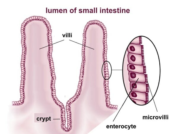
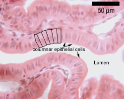
Respiratory system: Anatomy and functions - Kenhub 16-02-2022 · Respiratory system (Systema respiratorum) The respiratory system, also called the pulmonary system, consists of several organs that function as a whole to oxygenate the body through the process of respiration (breathing).This process involves inhaling air and conducting it to the lungs where gas exchange occurs, in which oxygen is extracted from the air, and carbon … Important Questions for CBSE Class 9 Science Chapter 6 ... Free PDF download of Important Questions with solutions for CBSE Class 9 Science Chapter 6 - Tissues prepared by expert Science teachers from latest edition of CBSE(NCERT) books. Register online for Science tuition on Vedantu.com to score more marks in your examination. Simple columnar epithelium Simple columnar epithelium (40X) Primate small intestine You are looking at part of the small intestine. The inner surface of the intestinal wall is made of simple columnar epithelium (sce). Instead of being smooth, the inside of the intestine is folded and covered by millions of tiny projections called villi. On the image, you can see one fold ... Simple Columnar Epithelium Diagram - Quizlet The surface area is increased with microvilli and its function is to move materials, secrete fluids, and absorb nutrients. What are goblet cells and their functions? Goblet cells are glandular simple columnar epithelial cells whose function is to produce and secrete mucus in order to protect the mucous membranes where they are found.
Biology Edited by Dr. Linda Sallah Fawzi
Epithelial Tissue: Structure with Diagram, Function, Types ... Columnar- long or column-like cylindrical cells, which have nucleus present at the base. On the basis of the number of layers present, epithelial tissue is divided into the simple epithelium and stratified or compound epithelium. Simple Epithelium- it is composed of one layer of a cell and mostly has a secretory or an absorptive function

Transitional Epithelium Functions, Location and Diagram ...
Ch. 5 LearnSmart Flashcards - Quizlet Start studying Ch. 5 LearnSmart. Learn vocabulary, terms, and more with flashcards, games, and other study tools.

Simple Squamous Epithelium Diagram | Quizlet
Epithelial Tissue | Anatomy and Physiology I Like the cuboidal epithelia, this epithelium is active in the absorption and secretion of molecules. Simple columnar epithelium forms the lining of some sections of the digestive system and parts of the female reproductive tract. Ciliated columnar epithelium is composed of simple columnar epithelial cells with cilia on their apical surfaces.

Epithelium Lab
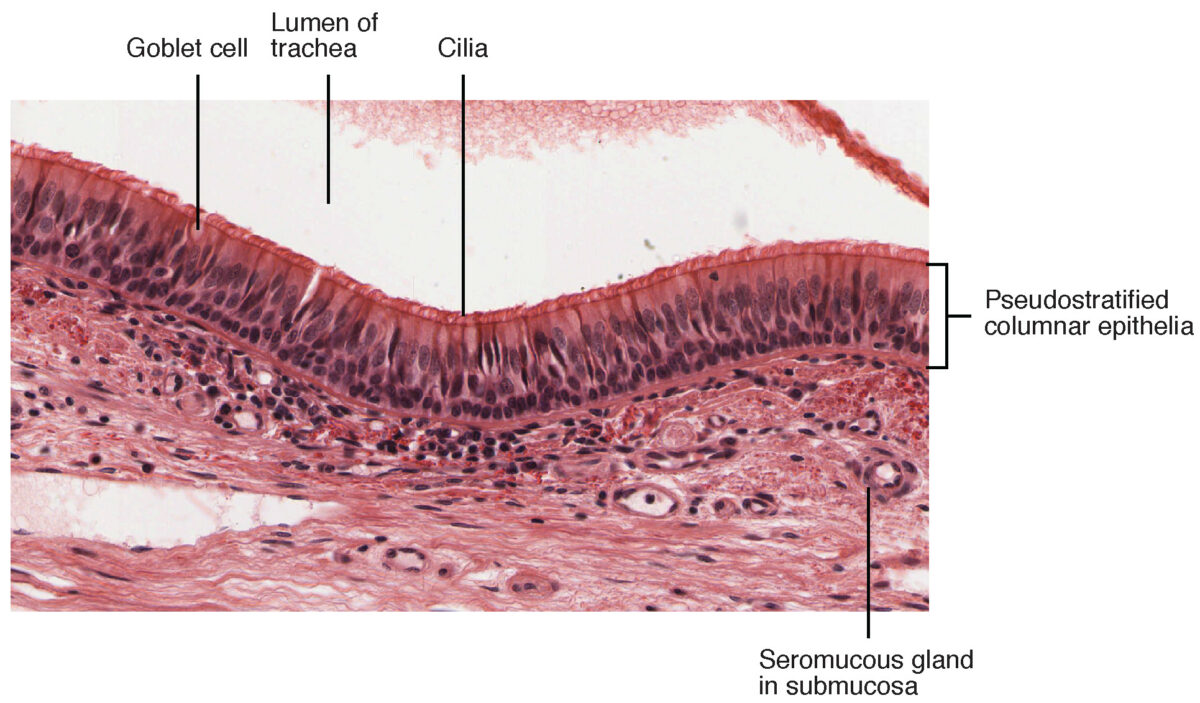
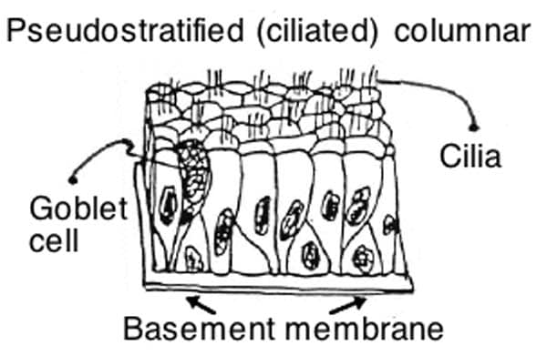
Epithelia: The Histology Guide Simple secretory columnar epithelium lines the stomach and uterine cervix.The simple columnar epithelium that lines the intestine also contains a few goblet cells. In histological slides of pseudostratified epithelium, it looks as though some of the cells are not in contact with the basal lamina, and the nuclei are at different levels.

Simple Squamous Epithelium 40X - Annotated | Histology
Simple columnar epithelium- structure, functions, examples The simple columnar epithelium is a type of epithelium that is formed of a single layer of long, elongated cells mostly in areas where absorption and secretion are the main functions. Like cuboidal epithelium, the cells in the columnar epithelium are also modified to suit the function and structure of the organ better.

Simple Columnar Epithelium - an overview | ScienceDirect Topics
Simple Squamous Epithelium: Location and Diagram - Video ... The simple squamous epithelium is different from other types of epithelial tissue such as simple cuboidal, simple columnar, and stratified squamous epithelium in that it is only made of one layer ...

Study Notes
Ileum Histology Slide with Labeled Diagram and ... I think the features mentioned earlier in the ileum histology slide labeled diagrams might help you a lot. Again, I will show you the ileum histology diagram where you might identify the following important features. ... The surface epithelium (simple columnar epithelium) contains microvilli on their surfaces. You may also find the goblet cells ...
Ciliated Columnar Epithelium
Simple Cuboidal Epithelium Function & Location | What Is ... Simple Cuboidal Epithelium: Labeled Diagram. ... In terms of structure, it is only the simple columnar, cuboidal, and squamous epithelium that is made up of a single layer of cells.
Simple Columnar Epithelium - Definition & Function | Biology ...
Simple Columnar Epithelium - Definition & Function ... Simple columnar epithelia are tissues made of a single layer of long epithelial cells that are often seen in regions where absorption and secretion are important features. The cells of this epithelium are arranged in a neat row with the nuclei at the same level, near the basal end. In a cross-section of the organ, these cells appear like thin ...

Simple Squamous Epithelium Function, Location, Structure and ...
Simple Columnar Epithelium Labeled Diagram Simple Columnar Epithelium: A Labeled Diagram and Functions Epithelium is a tissue that lines the internal surface of the body, as well as the internal organs. Simple epithelium is one of the types of epithelium that is divided into simple columnar epithelium, simple squamous epithelium, and simple cuboidal epithelium.

Epithelium Lab
Simple columnar epithelium - Eugraph These absorptive cells are a single layer of columnar cells. (a simple columnar epithelium). Note an oval nucleus in the lower part of each columnar cell. Arrows indicate the base of this simple columnar epithelium sce. The lumen is indicated by lu. The surface area for absorption is increased by projections of the intestinal wall called villi.

Pseudostratified columnar epithelium - Wikipedia
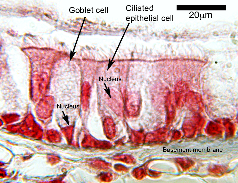
PDF Basic Histo diagrams labelled in colour - 2005 These labelled diagrams should closely follow the current Science courses in histology, anatomy and ... EPITHELIUM: simple columnar TISSUE / ORGAN: gall bladder EPITHELIUM: pseudostratified columnar (respiratory epithelium) MORE FULLY: pseudostratified ciliated columnar epithelium with goblet cells TISSUE / ORGAN: trachea EPITHELIUM:
Squamous Epithelium High Resolution Stock Photography and ...
Stomach histology: Mucosa, glands and layers - Kenhub The surface epithelium is a simple columnar epithelium. It lines the inside of the stomach as surface mucous cells and forms numerous tiny invaginations, or gastric pits, which appear as millions of holes all throughout the stomach lining. These gastric pits are important as they are connected to the various glands of the stomach.

57 Ciliated Epithelium Photos and Premium High Res Pictures ...
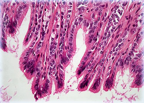
Epithelial Tissue | Boundless Anatomy and Physiology Simple columnar epithelium is a single row of tall, closely packed cells, aligned in a row. These cells are found in areas with high secretory function (such as the wall of the stomach), or absorptive areas (as in small intestine ).

Simple Squamous Epithelium
Tooth development – Histology and Embryology for Dental ... Figure 8.5: Animated overview of the early stages of tooth development: Placode, bud, cap, bell. Bud stage. Continued proliferation of the ectoderm allows us to see the next stage of tooth development under the microscope more easily, the bud stage.The name bud comes from the fact that tooth buds look like leaf buds on a plant. In the spring, you can see where leaves are …

Epithelial Tissues | Biology for Majors II
Epithelial Tissue - Anatomy and Physiology Like the cuboidal epithelia, this epithelium is active in the absorption and secretion of molecules. Simple columnar epithelium forms the lining of some sections of the digestive system and parts of the female reproductive tract. Ciliated columnar epithelium is composed of simple columnar epithelial cells with cilia on their apical surfaces.

Simple Squamous Epithelium - ppt video online download
PDF Marieb HA8 chapter 4 - Pearson cells. This epithelium forms the secretory cells of many glands, the walls of the smallest ducts of glands, and the walls of many tubules in the kidney. Its functions are the same as those of simple columnar epithelium. Simple Columnar Epithelium (Figure 4.3c) Simple columnar epithelium is a single layer of tall cells aligned like soldiers in a ...

Simple Columnar Epithelium
Simple Columnar Epithelium Labeled Diagram Simple Columnar Epithelium Labeled Diagram Ciliated columnar epithelium is composed of simple columnar epithelial cells with cilia on their apical This illustration shows a diagram of a goblet cell. These labelled diagrams should closely follow the current Science (simple squamous epithelium). ORIGIN: columnar epithelium with goblet cells. TISSUE .

Pseudostratified Columnar Epithelium Function & Location ...

Lab 2 Epithelial tissue | Histology
Epithelial Tissues Stock Illustrations – 30 Epithelial ...

Simple Columnar Epithelium Labeled Diagram and Function
Simple columnar epithelium Images, Stock Photos & Vectors ...

Specialisation of Cells | Luna Perez Muñiz

simple squamous epithelium - Google Search | Histology slides ...
Simple epithelium: Location, function, structure | Kenhub
Surface Epithelium | Concise Medical Knowledge
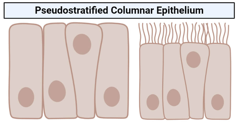
Pseudostratified columnar epithelium- structure, functions ...
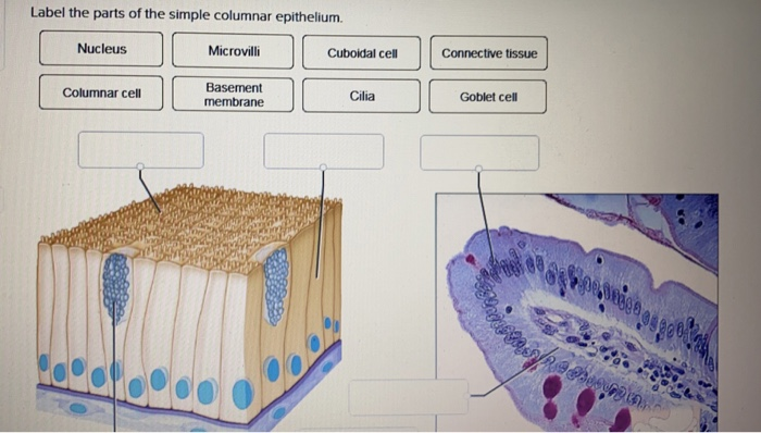
Solved Label the parts of the simple columnar epithelium ...

Describe Various Types of Epithelial Tissues with the Help of ...
Epithelia: The Histology Guide

Simple columnar epithelium Simple squamous epithelium ...
Simple Columnar Epithelium | Olympus LS
(253).jpg)
Epithelial Tissue Quiz - ProProfs Quiz
Simple Columnar Epithelium: A Labeled Diagram and Functions ...
Pseudostratified Columnar Epithelium | Histology, Anatomy & Types

Simple squamous epithelium

How to draw stratified columnar epithelium || easy way
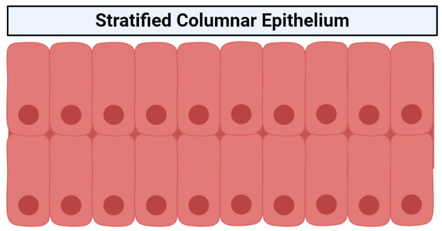
Stratified columnar epithelium- structure, functions, examples
Tissue Review Slides for Human Anatomy

Exercise 4: Epithelium
Where is the location and function of an epithelial tissue ...
Epithelia: The Histology Guide
0 Response to "43 simple columnar epithelium labeled diagram"
Post a Comment