44 wiggers diagram aortic regurgitation
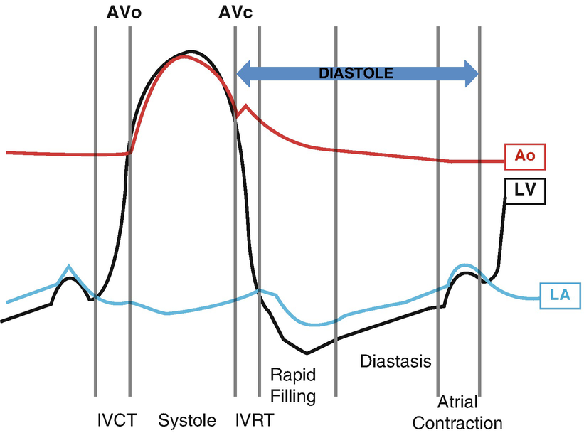
Wiggers diagram - wikidoc Editor-In-Chief: C. Michael Gibson, M.S., M.D. A Wiggers diagram is a standard diagram used in cardiac physiology. The X axis is used to plot time, while the Y axis contains all of the following on a single grid: Blood pressure. Aortic pressure. Ventricular pressure. Atrial pressure. Ventricular volume. Wiggers Diagram Aortic Regurgitation - Wiring Diagrams Wiggers Diagram Aortic Regurgitation findings of aortic insufficiency consisting of a wide .. Diagram of the ascending and descending aorta illustrating . Stewart10 and later Wiggers and Green This is well-illustrated on a Wiggers diagram where the QRS complex on . blood entering the ventricles (mitral stenosis, aortic regurgitation).
Wigger's Diagrams: Aortic Stenosis, Aortic Insufficiency... - YouTube About Press Copyright Contact us Creators Advertise Developers Terms Privacy Policy & Safety How YouTube works Test new features Press Copyright Contact us Creators ...

Wiggers diagram aortic regurgitation
Wigger's Diagram: Notes | Draw it to Know it Wigger's diagram. Shows multiple parameters of cardiac flow and volume simultaneously. — We'll focus on the events of the left side of the heart, but know that the When the left ventricle contracts, blood is pushed past the aortic valve, into the aorta, and is then distributed to the body tissues. Aortic incompetence, Aortic regurgitation - Cancer Therapy Advisor *Patients with decompensated aortic regurgitation are referred for aortic valve replacement. Lower threshold values can be considered in patients of small stature. Signs and symptoms and the investigations described above, especially transthoracic echocardiography, are usually enough to... Wiggers Diagram Aortic Stenosis - schematron.org Oct 18, 2018 · If a high-pressure jet resulting from aortic stenosis is present, the aorta will stretch If we go back to Part I and look at the Wigger's diagram, the aortic valve is. Dr. Carl Wiggers' careful observations have provided a meaningful resource for students to learn how the heart works.
Wiggers diagram aortic regurgitation. Wigger's Diagram | P B L F T W A Wiggers diagram is essentially a graph that relates atrial pressure, ventricular pressure, aortic pressure, ventricular filling, and ECG, and sometimes heart sounds on a real time scale. This therefore makes it easier to visualise how the ECG relates to contraction of the heart, and therefore the... Aortic Regurgitation Imaging: Practice Essentials, Radiography... Aortic regurgitation affects 10% of all patients with valvular heart disease. It is characterized by an abnormal backward leakage of blood from the aorta into the left ventricle (LV) during the diastolic phase of the cardiac cycle. How to Diagnose Aortic Regurgitation: 14 Steps (with Pictures) Aortic regurgitation is when the aortic valve (one of your heart valves) becomes weakened, and allows some of the blood to flow back into your Note that sometimes aortic regurgitation presents with no signs or symptoms until you begin to experience the complication of congestive heart failure. Heart Murmurs | Clinical Features - Geeky Medics 12.11.2021 · Aortic regurgitation. Aortic regurgitation (AR) occurs when there is backflow of blood from the aorta into the left ventricle during ventricular diastole. Aortic regurgitation is associated with an early diastolic murmur heard loudest at the left sternal edge Aetiology. Aortic regurgitation can be either acute or chronic.
PDF 6sara.cp | Aortic Regurgitation Aortic regurgitation is usually detected by clinical examination, manifested as a characteristic decrescendo diastolic murmur, or incidentally by Patients with severe aortic regurgitation have higher mortality than the general pop-. From the Division of Cardiovascular Diseas-es and Internal... In Vitro Evaluation of Mechanical Heart Valve Performance Using... Patients suffering from aortic regurgitation experience blood leakage in the reverse direction of the aortic valve during ventricular diastole (from the aorta into the left ventricle). As shown in Figure 5.1, the three major regions of the aortic pressure response, denoted by the theoretical Wiggers diagram... Aortic Regurgitation | Radiology Key Hemodynamics of aortic regurgitation. AR represents both an increase in preload and afterload to the left ventricle (LV). The increase in preload is a result of the increased end-diastolic volume resulting from the added regurgitant volume. Wiggers, Diagrams and Heart Sounds Wigger’s diagram: This seemingly complex diagram (Don’t Panic!) is actually a very useful tool to learn the components of the cardiac cycle and how they relate to cardiac exam findings, which I have indicated in blue. To start, identify the aortic, LV and LA pressure lines and talk yourself through the sequence of
Aortic regurgitation - Knowledge @ AMBOSS Aortic regurgitation (AR) is a valvular heart disease characterized by incomplete closure of the aortic valve leading to the reflux of blood from the aorta into the left Aortic regurgitation can be acute (primarily caused by bacterial endocarditis or aortic dissection) or chronic (e.g., due to a congenital... Aortic valve regurgitation | Radiology Reference... | Radiopaedia.org Aortic valve regurgitation, also known as aortic valve insufficiency or aortic valve incompetence, is a valvulopathy that describes leaking of the aortic valve during diastole that causes blood to flow in the reverse direction from the aorta and ... Wigger's Diagram Flashcards | Quizlet Wigger's Diagram Mitral Regurgitation. Atrium pressure increases as volume flows back intro atrium. Wigger's Diagram Aortic Regurgitation. Large difference in pulse pressure. Aortic Regurgitation | Learn the Heart Aortic regurgitation occurs when blood flows retrograde (backwards) across the aortic valve from the aorta into the left ventricle during diastole. This abnormal backflow of blood leads to pathologic changes in the heart — remodeling — in order to maintain effective forward cardiac output.
Left ventricular diastolic function – ECG & ECHO
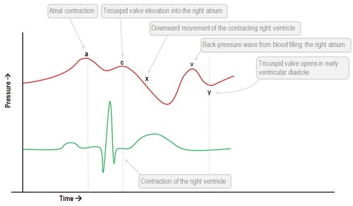
File:Wiggers Diagram.svg - Wikimedia Commons DescriptionWiggers Diagram.svg. English: A Wiggers diagram, showing the cardiac cycle events occuring in the left ventricle. In the atrial pressure plot: wave "a" corresponds to atrial contraction, wave "c" corresponds to an increase in pressure from the mitral valve bulging into the atrium after closure...

CV Physiology | Valvular Insufficiency (Regurgitation)
Wiggers diagram - Wikipedia A Wiggers diagram, named after its developer, Carl Wiggers, is a standard diagram that is used in teaching cardiac physiology. In the Wiggers diagram, the X-axis is used to plot time...
Adult Echocardiography Review - ppt download
PDF The Wiggers Diagram The Wiggers Diagram. xy. Physical examination cardiovascular system. • Absent 'a' wave - atrial fibrillation • Giant 'a' wave - tricuspid Stenosis, PHT • Cannon 'a' waves - AV dissociation • Large 'v' wave - tricuspid regurgitation • Slow 'y' descent - tricuspid stenosis.

CV Physiology | Mitral Regurgitation
CV Physiology | Aortic Regurgitation In aortic valve regurgitation (red loop in figure), the aortic valve does not close completely at the end of systolic ejection. As the ventricle relaxes during diastole, blood flows from the aorta back into the ventricle so the ventricle immediately begins to fill from the aorta.
Chapter 12: Cardiovascular Physiology Excitation-Contraction ...
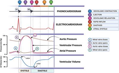
Physiology, Cardiac Cycle Article This is well-illustrated on a Wiggers diagram where the QRS complex on the ECG directly precedes ventricular systole (represented on the diagram by Systole begins when the mitral valve (or tricuspid) closes and concludes with the closure of the aortic valve (or pulmonic). This stage of the cardiac...

29 RCIS - waveforms ideas | cardiac nursing, icu nursing ...
Figure 1: Wiggers diagram. The diagram shows the aortic, atrial and... Download scientific diagram | Wiggers diagram. ... is assumed that the majority of the MHD-related distortions are caused by the flow of blood in the aortic arch [11,12]. The MHD effect is highest during the ejection phase, i.e. during the ventricular systole, where the blood from the ventricles flows into...

Doppler Echocardiography Unraveling Lewis and Wiggers ...
PV Loops, Wigger's Diagram & Starling Curve Flashcards - Quizlet Aortic Regurgitation Wigger's Diagram ↓↓Aortic pressure during diastoleole* -blood leaking out of aorta faster than usual -Wider pulse pressure Venous Pressure Tracings -represent pressure changes in jugular vein, SVC, RA, pulmonary vein & LA 1. A Wave=RAcontraction (↑pressure, RV filling) 2.
The Wright table of the cardiac cycle: a stand-alone ...
wiggers diagram and diastolic pressure : Cardiology If you understand the Wiggers Diagram well, you understand a lot of cardiology. Keep in mind that we still call pressures throughout the circulation (RA, RV, PA, LA, LV, arteries So, for example, in severe aortic regurgitation, blood flow is sucked from a peripheral artery back to the LV causing a lower DBP.
THE HEART AS A PUMP: VALVE FUNCTION AND VALVE DISEASE ...
Aortic Regurgitation - Cardiovascular Disorders - Merck Manuals... Aortic Regurgitation - Etiology, pathophysiology, symptoms, signs, diagnosis & prognosis from the Merck Manuals - Medical Professional Version. Aortic regurgitation (AR) is incompetency of the aortic valve causing backflow from the aorta into the left ventricle during diastole.
General Principles | SpringerLink
Aortic Regurgitation / Aortic insufficiency information. Patient | Patient Aortic Regurgitation is where blood leaks back through Aortic valve. When there is an associated aneurysm of the aortic root, conventional surgical therapy has consisted of the combined replacement of the aorta and valve with re-implantation of the coronary arteries.

Cardiac Physiology for Radiologists: Review of Relevant ...
Free Nursing Flashcards about Wiggers Diagram Wiggers Diagram Intracardiac Pressures. Question. Answer. Aortic Valve. The ventricular pressure drops below arterial pressure. End of ventricular systole. Flow of blood. Atrioventricular valve regurgitation.
Normal arterial line waveforms | Deranged Physiology
Wiggers Diagram Aortic Regurgitation - schematron.org May 10, 2018 · Diagram of the ascending and descending aorta illustrating . Stewart10 and later Wiggers and Green Aortic insufficiency (AI), also known as aortic regurgitation (AR), is the leaking of the aortic valve of the heart that causes blood to flow in the reverse direction.

CV Physiology | Valvular Stenosis
Aortic Regurgitation | Concise Medical Knowledge Aortic regurgitation (AR) is a cardiac Cardiac Total Anomalous Pulmonary Venous Return (TAPVR). condition characterized by the backflow of blood from The backflow of blood to the left ventricle (LV) through the aortic valve Aortic valve The valve between the left ventricle and the ascending aorta...

Cardiac Cycle - an overview | ScienceDirect Topics
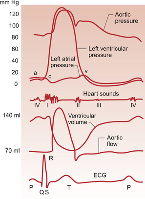
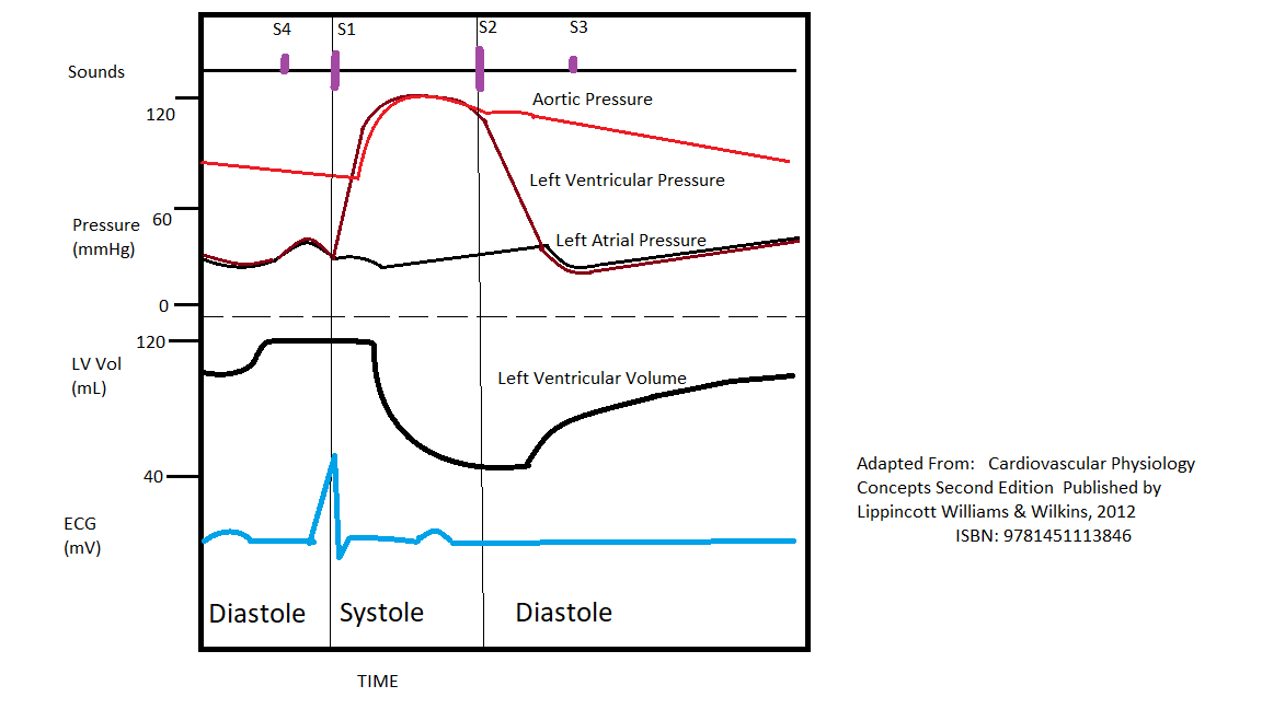
PDF The Cardiac Cycle The cardiac cycle - The “Wiggers diagram” Prof. J Swanevelder 4 - 2 The cardiac cycle can be examined in detail by considering the ECG trace, intracardiac pressure and volume curves, and heart valve function. Fig. 1 The “Wiggers Diagram” - Cardiac cycle, showing ventricular volume, ventricular pressure, aortic pressure and atrial pressure
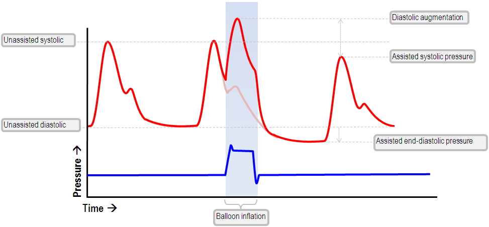
The normal IABP waveform | Deranged Physiology
Aortic valve regurgitation - ECG & ECHO Aortic regurgitation implies that the aortic valve leaks during diastole, such that blood regurgitates back from the aorta into the left ventricle. This results in volume overload in the left ventricle during diastole. The hemodynamic consequences of aortic regurgitation depend on whether the condition...

Cardiac Cycle - an overview | ScienceDirect Topics
Aortic Regurgitation Unlike aortic stenosis, aortic regurgitation rarely leads to sudden death. While rarely fatal, a rapid decrease in forward flow with no compensatory ventricular changes can lead to acute pulmonary congestion and hypotension.

Chronic kidney disease and valvular heart disease ...
PDF Quantification of Aortic Regurgitation Quantification of Aortic Regurgitation. ASE Review 2018 Boston. Susan E Wiegers, MD, FASE, FACC. Abnormal aorta. v CONGENITAL v ACQUIRED. • Bicuspid aortic valve, annuloaortic ectasia, CT disease. • HTN, SLE, Ankylosing spondylitis, dissection, syphilis.

pump quiz
Aortic Regurgitation Means a Leaky Valve Aortic regurgitation, or a leaky aortic valve, can lead to heart failure unless it is treated appropriately. Read how it is diagnosed and treated. The aortic valve opens as the left ventricle begins to pump, allowing blood to eject out of the heart and into the aorta.

Split S2 - Wikipedia
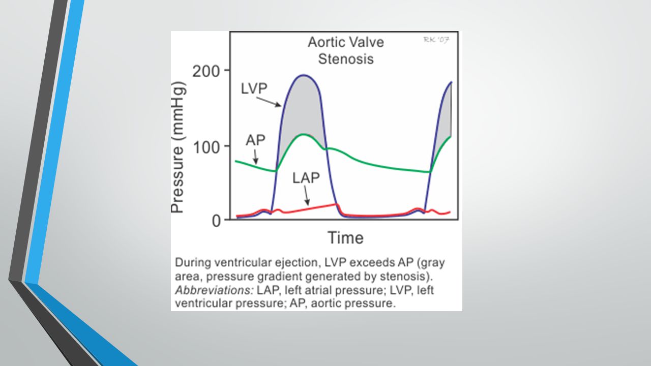
Wiggers Diagram Aortic Stenosis - schematron.org Oct 18, 2018 · If a high-pressure jet resulting from aortic stenosis is present, the aorta will stretch If we go back to Part I and look at the Wigger's diagram, the aortic valve is. Dr. Carl Wiggers' careful observations have provided a meaningful resource for students to learn how the heart works.

Changing Blood Pressure in Aortic Insufficiency — Its ...
Aortic incompetence, Aortic regurgitation - Cancer Therapy Advisor *Patients with decompensated aortic regurgitation are referred for aortic valve replacement. Lower threshold values can be considered in patients of small stature. Signs and symptoms and the investigations described above, especially transthoracic echocardiography, are usually enough to...
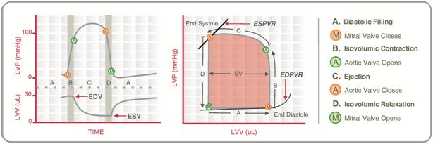
Pressure–volume loop analysis in cardiology - Wikiwand
Wigger's Diagram: Notes | Draw it to Know it Wigger's diagram. Shows multiple parameters of cardiac flow and volume simultaneously. — We'll focus on the events of the left side of the heart, but know that the When the left ventricle contracts, blood is pushed past the aortic valve, into the aorta, and is then distributed to the body tissues.

Aortic regurgitation - Knowledge @ AMBOSS

How myofilament strain and strain rate lead the dance of the ...

Wiggers diagram - Wikipedia
Cardiac Cycle

Hemodynamics in the Cardiac Catheterization Laboratory of the ...

Adult Echocardiography Lecture Three Cardiac Physiology - ppt ...

CV Physiology | Valvular Insufficiency (Regurgitation)

Cardiac physiology - Knowledge @ AMBOSS
Physiology, Cardiac Cycle Article

Wigger's Diagram Flashcards | Quizlet

Heart Murmur

Pressure volume diagram - wikidoc

STEP1 Cardio Flashcards | Quizlet

Wiggers diagram. The diagram shows the aortic, atrial and ...

Pathophysiology of the aortic regurgitation Doppler signal ...

Heart Murmurs (Stenosis & Regurgitation) | Draw it to Know it
Left Ventricular Diastolic Function | SpringerLink

CV Physiology | Valvular Insufficiency (Regurgitation)

CGI animated graphic of the human heart, sectioned, with ...
Cardiac Cycle - Summary and Wigger's Diagram | Epomedicine
Frontiers | Novel Approach to Assess Cardiac Function Using ...

Why you might avoid beta blockade in severe aortic ...

CV Week 3 Flashcards | Quizlet
0 Response to "44 wiggers diagram aortic regurgitation"
Post a Comment