41 dog muscle anatomy diagram
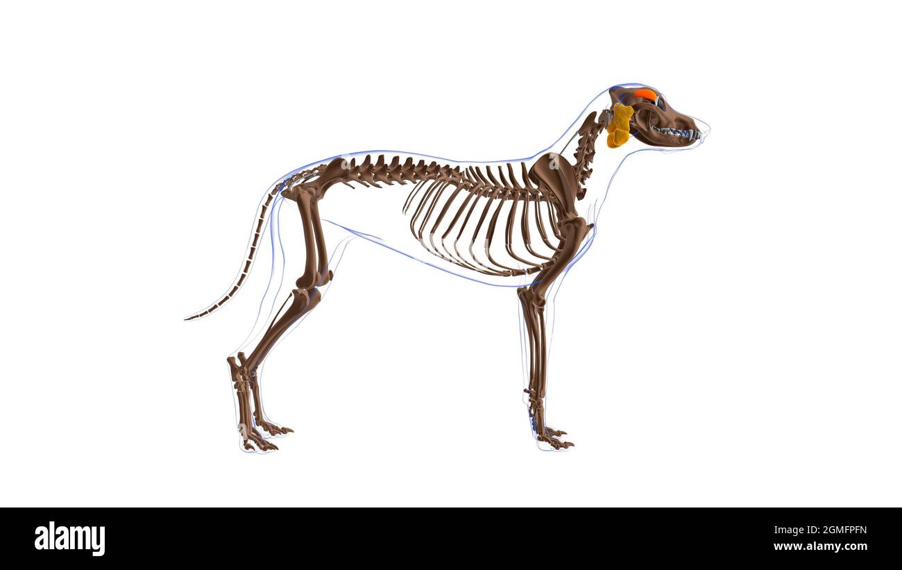
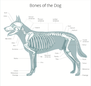
Dog Anatomy Guide - Muscles, Bones and Organs These all facilitate the attachment of various muscles in the dog's head. The dog's spine is divided into cervical vertebrae, thoracic vertebrae, lumbar vertebrae, sacral vertebrae and coccygeal vertebrae. Cervical vertebrae: there are seven cervical vertebrae in every dog, even if some dogs may have a longer neck than others. PDF Canine Muscle Origins, Insertions, Actions and Nerve ... Canine Muscle Origins, Insertions, Actions and Nerve Innervations The purpose of this document is to provide students of canine anatomy a simple reference for muscular origins, insertions, actions and nerve innervations without having to search through the overwhelming verbiage that accompanies most canine anatomy texts. Millerʼs Anatomy of
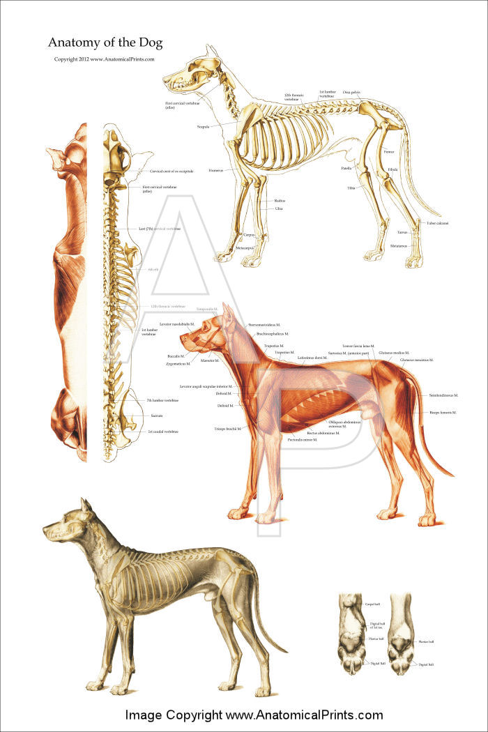
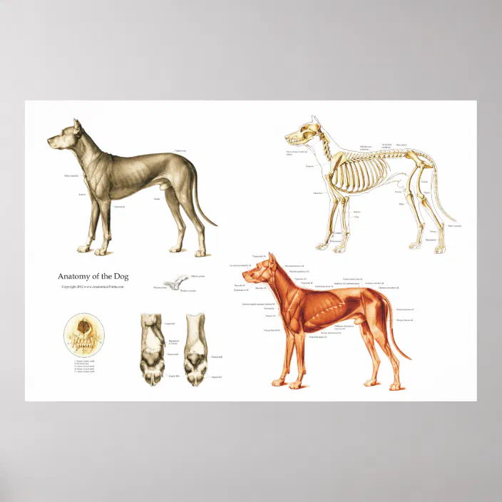
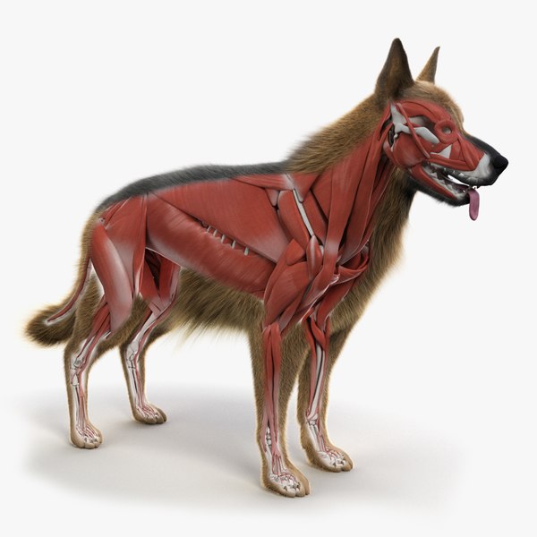
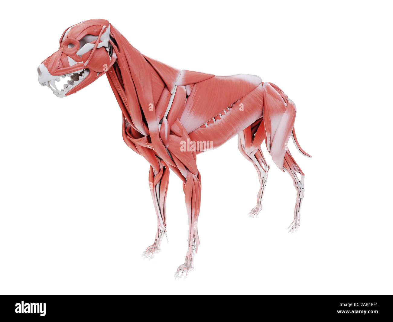
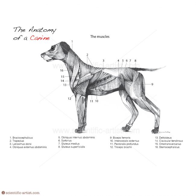
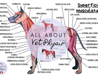
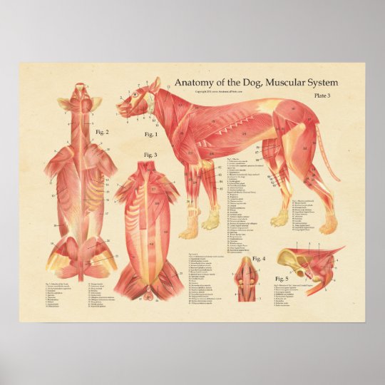
Dog Muscle Anatomy Diagram In this image, you will find scutularis, sternomastoideus, brachiocephalicus, trapezius, latissimus dorsi, iliocostalis, sartorius, tensor fasciae latae, glutaueus medius, crest of pelvis bone, glutaeus superficialis, trochanter major, semitendinosus, biceps femoris, quadriceps femoris, gastrocnemius, flexor hallucis longus in dog muscle anatomy.

Dog muscle anatomy diagram
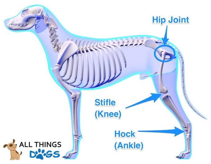
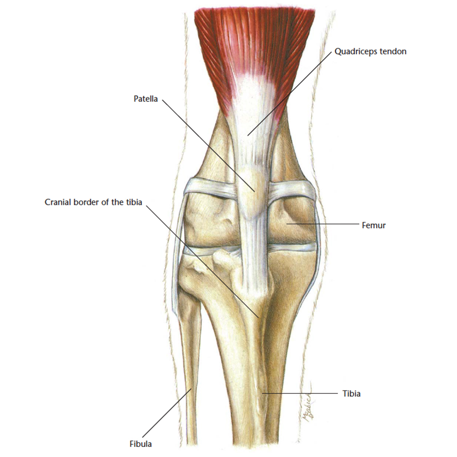
Dog Knee Anatomy with Labeled Diagram » AnatomyLearner ... "There are two menisci in the knee joint of a dog (showed in the diagrams)" The femoropatellar joint of dog knee The femoropatellar joint locates between the patella and trochlea of the femur. You will find two slightly oblique ridges with a wide and deep groove within them in the trochlea of a dog. 2021 Ultimate Veterinary Guide to Dog Anatomy with Images ... It is made up of skeletal bones, muscles, cartilage, tendons, ligaments, joints and connective tissue. Common joints include the: Elbow Shoulder Hip Stifle (knee) Shoulder The shoulder joint is made up of the scapula (shoulder blade) and humerus (large arm bone). VetCheck Passive Range of Motion Guidelines A Visual Guide to Understanding Dog Anatomy With Labeled ... The following diagram and paragraph attempt to explain it in brief. The muzzle is of varying lengths, depending on the breed. Whiskers, present on the muzzle, are of some sensory use. Dogs also have a 'stop' on their heads, which is the point where the muzzle ends and the forehead begins.
Dog muscle anatomy diagram. Dog Anatomy - Dog.com Dog Anatomy: Canine Physique - The Body of your Dog. Because dogs have been selectively bred for thousands of years, there are a tremendous variety of body types and sizes. ... The powerful muscles in a dog's hind legs give them an incredible ability to jump. Many can leap three times their own height. EasyAnatomy | Interactive 3D Canine Anatomy Software For Practitioners. Practitioners and their clients benefit from EasyAnatomy's interactive canine model and animations of common pathologies. As the most advanced interactive 3D canine anatomy client communication tool, EasyAnatomy breaks down the communication barrier and increases client compliance. Learn more. Dog Tongue Anatomy with Labeled Diagram - Muscles ... Dog Tongue Anatomy with Labeled Diagram - Muscles, Papillae, Glands, Veins, and Nerves 13/01/2022 12/01/2022 by anatomylearner The dog tongue anatomy consists primarily of skeletal muscle, mucous membrane, glands, vessels, and nerves. You will find different important anatomical facts on the mucous membrane of a dog tongue. Dog Spine Anatomy - Anatomical Features of Canine ... I hope the dog spinal nerve anatomy diagram might help you to understand all these branches so easily. You will find the spinal nerve in the cervical, thoracic, lumbar, sacral, and caudal regions of the dog anatomy. There are eight pairs of cervical spinal nerves present in the dog.
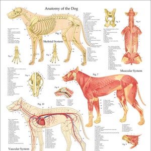
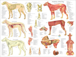
Dog Mouth Anatomy - Lip, Cheek, Oral cavity, and Salivary ... Dog lips anatomy The lips are the thick and rigid musculo-membranous structure surrounding the mouth orifice. Externally, it is formed by the skin, and internally it is lined by the pigmented epithelium. You will find the orbicularis oris muscle in between these two layers. Anatomy, medical imaging and e-learning for healthcare ... 301 Moved Permanently. nginx The Muscle Anatomy of Dogs - Everything You Need To Know The Muscle Anatomy of a Dog Pictured above is Ace from DarkDynastyK9s, arguably one of the most muscular dogs in the world. Ace's most predominant muscles are his triceps, Bicep Femoris, Scapular Deltoid, Acromion Deltoid, and his lower Trapezius. Ace uses Bully Max™ along with a 30/20 (30% Protein, 20% Fat) kibble. Dog Anatomy Posters and Charts - Anatomical Prints Dog Anatomy Muscles Poster 18" X 24" Shows the superficial and deep layer of muscles of the canine. Dog Skeletal Anatomy Poster 18" X 24" Shows the skeleton of the dog with special reference to the skull. Dog Anatomy Nerves Poster 18" X 24" Shows the nervous system, peripheral nerves and cranial nerves. Dog Vascular Anatomy Poster 18" X 24"
Dog Pelvis Anatomy - Male and Female Pelvic Limb Bone ... The lateral pelvis muscles of a dog include the followings - Tensor fasciae latae muscle of dog pelvis The gluteus superficial, medius, and profundus muscles of the dog pelvis, and Piriformis muscle of the dog pelvis Again, the medial pelvic muscles of the dog include obturator internus, obturator externus, Gemelli, and quadratus femoris muscle. The canine head and skull (CT): atlas of veterinary ... Labeled anatomy of the head and skull of the dog on CT imaging (bones of cranium, brain, face, paranasal sinus, muscles of head) This module of vet-Anatomy presents an atlas of the anatomy of the head of the dog on a CT. Images are available in 3 different planes (transverse, sagittal and dorsal), with two kind of contrast (bone and soft tissues). A Visual Guide to Dog Anatomy (Muscle, Organ & Skeletal ... Speaking of skeletons, a dog has 320 bones in their body (depending on the length of their tail) and around 700 muscles. Muscles attach to bones via tendons. Depending on the breed of dog, they will have different types of muscle fibers. You've probably heard about slow and fast twitch muscle fibers before. PDF CVM 6100 Veterinary Gross Anatomy In the diagram to the right if muscle #1 and muscle #2 both contract 10% during an identical time period - muscle #1's contraction would result in a larger movement of the lever arm during that same frame of time than muscle #2. In other words, muscle #1 will result in a more rapid rotation - it has a velocity advantage.
Dog Neck Anatomy - Bones, Muscle, Glands, Veins, and Other ... You know, there are seven cervical vertebrae in the dog skeleton (atlas, axis, third, fourth, fifth, sixth, and seventh cervical vertebrae). Again, the dog neck region's essential muscles are brachiocephalicus, omotransversarius, sternocephalicus, splenius, longus capitis, longus coli, scalenus, and serratus ventralis cervicis.
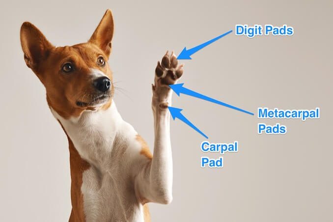
Dog Leg Anatomy with Labeled Diagram - Bones, Joints ... Now I will provide you the few information on the other bones of dog leg anatomy with their unique features. The front leg of a dog consists of the clavicle, scapula (arm), radius and ulna (forearm), carpals, metacarpals, and phalanges (forepaw). You will also find some palmar sesamoid bones in the front leg of a dog.
A Visual Guide to Understanding Dog Anatomy With Labeled ... The following diagram and paragraph attempt to explain it in brief. The muzzle is of varying lengths, depending on the breed. Whiskers, present on the muzzle, are of some sensory use. Dogs also have a 'stop' on their heads, which is the point where the muzzle ends and the forehead begins.
2021 Ultimate Veterinary Guide to Dog Anatomy with Images ... It is made up of skeletal bones, muscles, cartilage, tendons, ligaments, joints and connective tissue. Common joints include the: Elbow Shoulder Hip Stifle (knee) Shoulder The shoulder joint is made up of the scapula (shoulder blade) and humerus (large arm bone). VetCheck Passive Range of Motion Guidelines
Dog Knee Anatomy with Labeled Diagram » AnatomyLearner ... "There are two menisci in the knee joint of a dog (showed in the diagrams)" The femoropatellar joint of dog knee The femoropatellar joint locates between the patella and trochlea of the femur. You will find two slightly oblique ridges with a wide and deep groove within them in the trochlea of a dog.
















0 Response to "41 dog muscle anatomy diagram"
Post a Comment