43 hepatic portal vein diagram
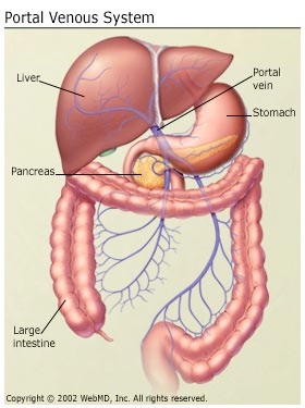
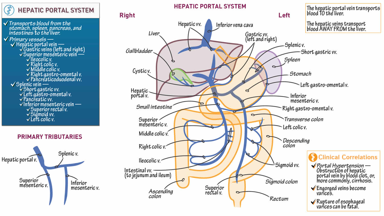
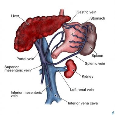
The portal venous system is part of the circulatory system. After the oxygenated blood from the hepatic vein and the blood from the portal vein mix in the sinusoids, it travels into a central vein and then drains into the hepatic vein. The mixed blood drains into the inferior vena cava. Through this pathway, the liver is provided with about 40 ... The hepatic portal vein is only about 3 inches (8 cm) long. It receives blood from the following blood vessels: 1) superior mesenteric vein - which collects blood from the stomach, small intestine and the ascending and transverse portion of the large intestine.
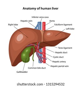
The liver has a dual blood supply, receiving most of its blood flow (75%) as deoxygenated blood from the portal vein, and the rest from the hepatic artery. The portal vein is a low-pressure system of valveless vessels which does not autoregulate according to hepatic oxygen demand, but rather according to supply (eg. with meals, the portal vein dilates and increases its flow).
Hepatic portal vein diagram
We are pleased to provide you with the picture named Hepatic portal system anatomical diagram.We hope this picture Hepatic portal system anatomical diagram can help you study and research. for more anatomy content please follow us and visit our website: www.anatomynote.com. Anatomynote.com found Hepatic portal system anatomical diagram from plenty of anatomical pictures on the internet. We are pleased to provide you with the picture named Portal Vein And Hepatic Vein In Liver.We hope this picture Portal Vein And Hepatic Vein In Liver can help you study and research. for more anatomy content please follow us and visit our website: www.anatomynote.com. Anatomynote.com found Portal Vein And Hepatic Vein In Liver from plenty of anatomical pictures on the internet. During hepatic resection, need for complete control of the hepatic vascular inflow may be accomplished by a Pringle maneuver. 5,6 This maneuver, developed by an Australian surgeon, James Hogarth Pringle, while in Glasgow, Scotland, during the management of hepatic trauma, involves occlusion of the hepatic artery and portal vein inflow through ...
Hepatic portal vein diagram. failure, tricuspid regurgitation, hepatic vein/portal vein fistula, or portal hypertension (Fig. 5). The flow in the splenic and superior mesenteric veins is toward (heptopedal) the liver, and both exhibit a low-veloc-ity, monophasic signal. Hepatic artery flow is in the same direction as the portal vein (hepatopetal). The hepatic artery Hepatic Portal System: In human anatomy, the hepatic system is the system is that the system of veins comprising the hepatic portal vein and its tributaries. Rat Liver Sinusoid: Sinusoid of a rat liver with fenestrated epithelial tissue cells. therefore, the Fenestrae area unit approx 100nm diameter and curved dimension five(5) microns. Start studying LABEL VEINS OF HEPATIC PORTAL SYSTEM. Learn vocabulary, terms, and more with flashcards, games, and other study tools. The porta hepatis is a transverse slit in the hilum of the liver that is perforated by the right and left hepatic ducts, hepatic artery, and portal vein. The common bile duct, hepatic artery, portal vein, nerves of the liver, and lymphatics lie enclosed within the layers of the hepatoduodenal ligament (free edge of lesser omentum).
On normal anatomy, typically, the splenic vein (SV) joins the superior mesenteric vein (SMV) anteriorly to the IVC and posteriorly to the pancreatic neck to form the PV, which ascends within the hepatoduodenal ligament, posteriorly to the hepatic artery and common bile duct, toward the hepatic hilum, where it divides into right and left (Fig. 2 a). Hepatic portal vein Renal vein Inferior mesenteric vein Inferior vena cava Vertebral vein External jugular vein Internal iliac vein . Term List (10) Radial vein . Axillary vein . Basilic vein . Brachial vein . Median cubital vein . Dorsal venous arch . Small saphenous vein . Digital veins . hepatic artery (black arrow), and main portal vein (arrowhead) are transmitted through the porta hepatis. MR imaging is often used as a problem-solving tool because of its superior soft-tissue resolution (Table 2). Bile in the common he-patic duct gives rise to its high signal intensity on MR images. The portal vein and hepatic Start studying hepatic portal vein. Learn vocabulary, terms, and more with flashcards, games, and other study tools.
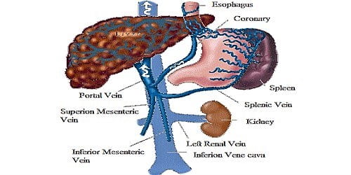
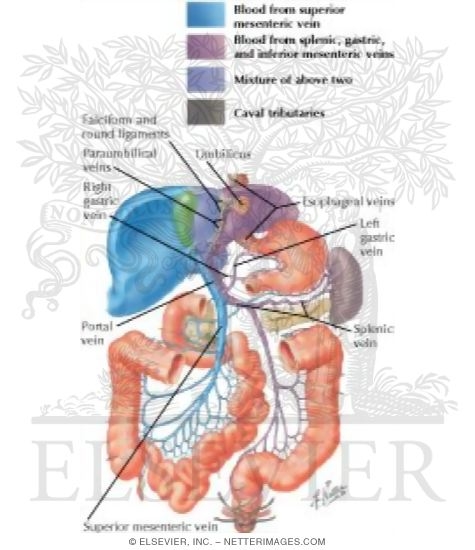
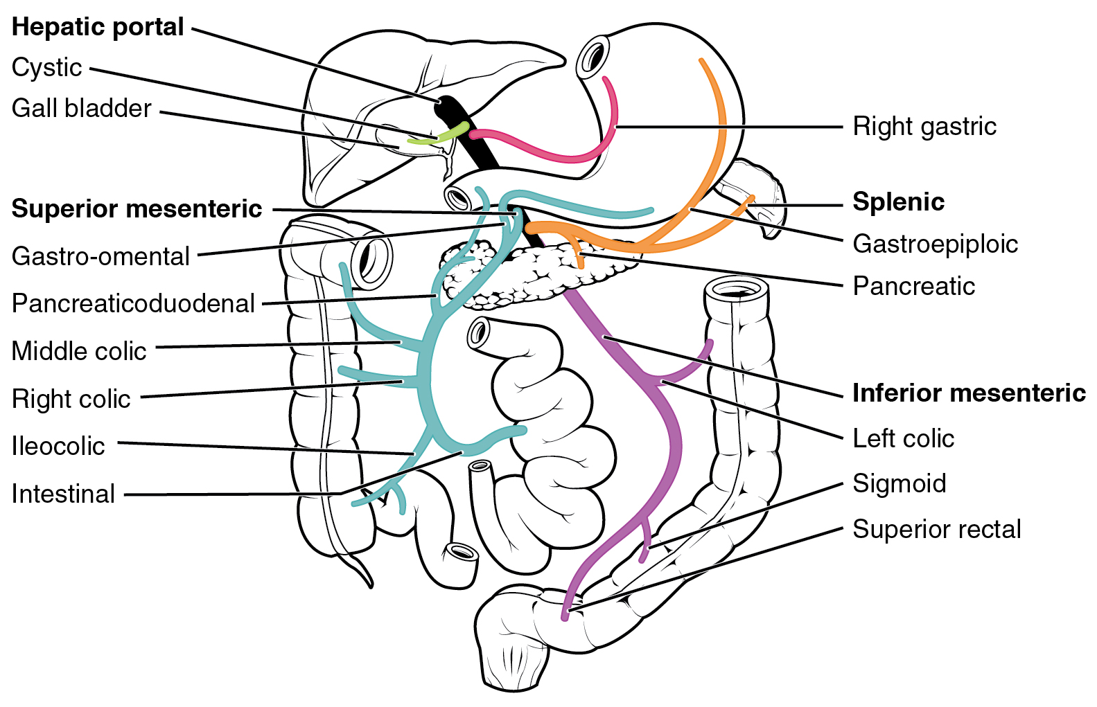
The porta hepatis is the central intraperitoneal fissure of the liver that separates the caudate and the quadrate lobes. It is the entrance and the exit for several important vessels including the portal vein, the hepatic artery, the hepatic nervous plexus, the hepatic ducts and the lymphatic vessels. The hepatic portal system is designed to require the digested elements to pass through the liver before entering the general circulation. The digestive organs are then drained by the hepatic portal vein. This vein is created by the conjuncture of the superior mesenteric vein and the splenic vein. HEPATIC PORTAL SYSTEM DIAGRAM The hepatic portal vein is the largest vein in the abdominal cavity. It drains blood from the spleen and the gastrointestinal tract to the liver. The hepatic veins begin at the junction of splenic veins and superior mesenteric. The blood from the cystic veins and the inferior mesenteric gastric veins is also drained by the hepatic vein. The hepatic veins drain directly into the inferior vena cava which feeds into the right atrium of the heart. From there the heart circulates the venous blood to the lungs to become oxygenated before it is sent back to the peripheral tissues. Portal Hypertension: Portal hypertension is an increase in the pressure of the portal vein.
Who Int
Portal vein. The hepatic portal vein is a vessel that moves blood from the spleen and gastrointestinal tract to the liver. It is approximately three to four inches in length and is usually formed ...
Differences Between Hepatic Portal System And Renal Portal System Qs Study
Portal pressure decreases from 09:00 to about 19:00 hours. Interestingly, peak reports of bleeding varicies correspond with 09:00 and 23:00 hours.* Normal hepatic circulation is a high flow - low resistance system. Branches of the portal vein deliver 1000-1500 ml/min of blood into the sinusoids of the hepatic lobules. Normal portal venous ...
Hepatic Portal System Images Stock Photos Vectors Shutterstock
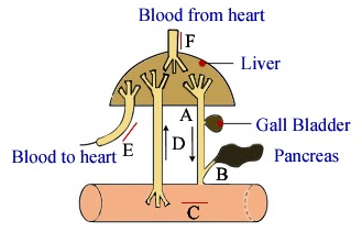
Following is a diagram showing the blood circulation of the body. We can see hepatic portal vein here (a part of portal system), this vein begins in a capillary (of intestines) and ends in another capillary (of liver). The hepatic portal circulation travels from the intestine of the digestive tract to the liver.

Hepatic Portal Veins Diagram Quizlet
In the hepatic portal system, the liver receives a dual blood supply from the hepatic portal vein and hepatic arteries. The hepatic portal vein carries venous blood drained from the spleen, gastrointestinal tract and its associated organs; it supplies approximately 75% of the liver's blood. The hepatic arteries supply arterial blood to the ...
Portal Venous Anatomy
The hepatic portal system consists of: Hepatic portal vein: This is the main vein connected to the liver.It forms at the connection of the inferior and superior mesenteric veins. Inferior ...

Hepatic Portal Vein An Overview Sciencedirect Topics
Download scientific diagram | Diagram of the hepatic segments (I-VIII) with their portal venous branches, separated by the hepatic veins and the transverse fissure. Anterior view of the liver.

Pin On Coc
The hepatic portal vein is one of the most important vein that receives blood from the body and transports it into the liver for filtration and processing. This vein is part of the hepatic portal system that receives all of the blood draining from the abdominal digestive tract, as well as from the pancreas, gallbladder, and spleen. 'Hepatic' means of or relating to the liver, therefore the ...
1
The main vessel of the hepatic portal system is the hepatic portal vein (Figures 3.31 and 3.32), a large vein that lies in the gastrohepatoduodenal ligament alongside the hepatic artery and anterior part of the bile duct.The hepatic portal vein is formed by the confluence of three main vessels, the gastric, pancreaticomesenteric, and lienomesenteric veins.

Anatomy Of Splenic Circulation And Placement Of Ligatures On Hepatic Download Scientific Diagram
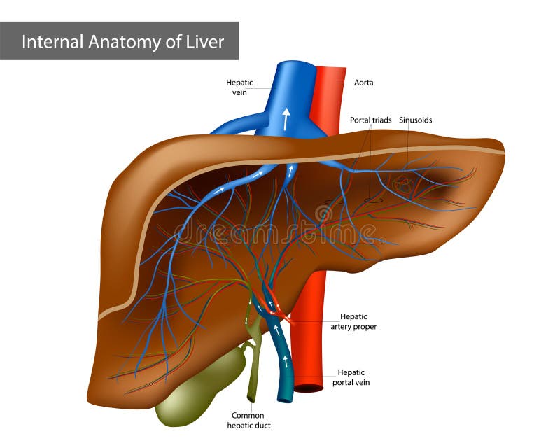
The main portal vein divides into the left portal vein, which supplies blood to the left lobe of the liver and the right portal vein, which supplies the right lobe of the liver . Portal veins characteristically run in triads with the hepatic arteries and bile ducts and branch away from the porta hepatis.
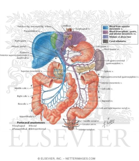
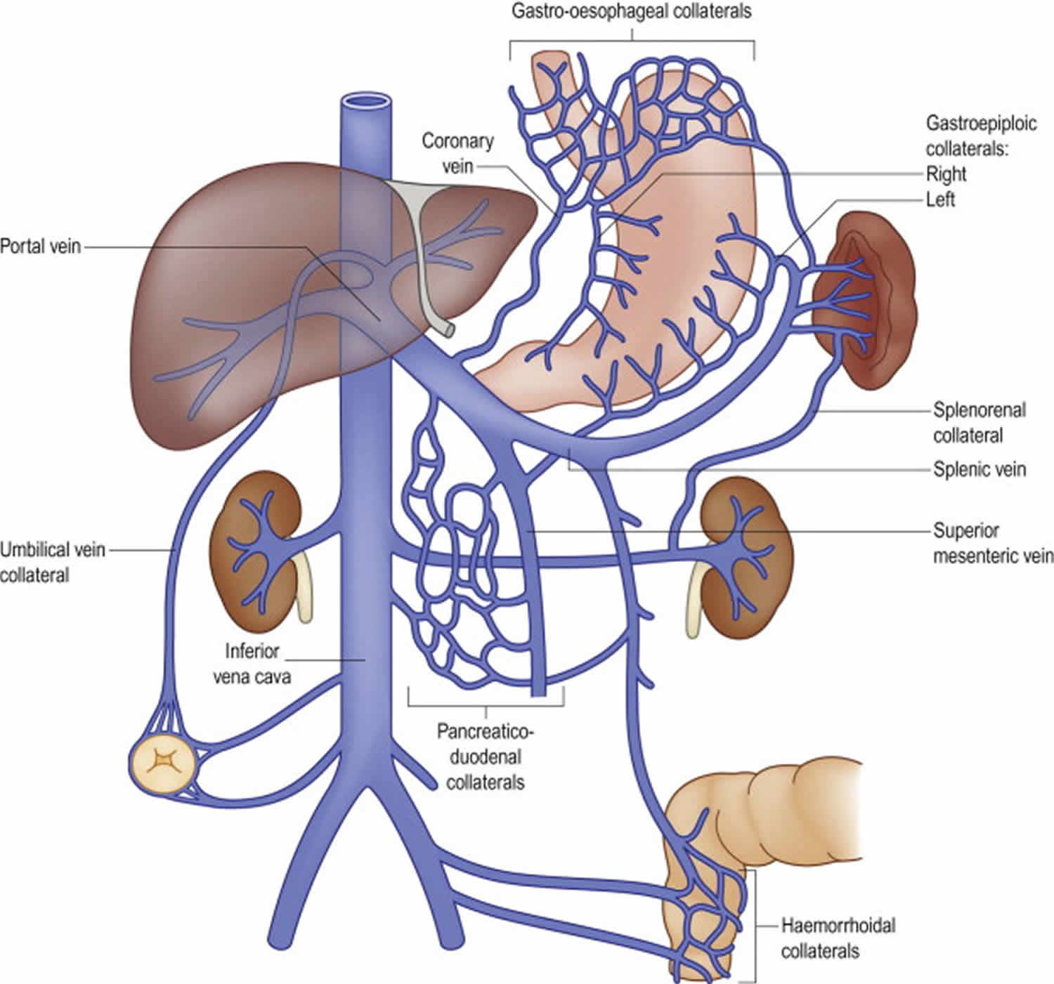
Hepatic Portal Vein Tributaries Portocaval Anastomoses
The renal portal vein receives the blood from the body wall of lumbar region through dorso-lumbar vein. Hepatic Portal System: Hepatic portal system collects the blood from the alimentary canal through many branches and carries it to the liver where the veins break up into capillaries and the blood is collected by hepatic veins to pour into the ...

0808 S2 Article 16
Left hepatic lobectomy involves removal of segments 2 to 4 and requires division of the left hepatic artery and left portal vein (inflow structures) as well the left hepatic duct and left hepatic vein (outflow structures). In a patient with a healthy background liver, anatomic trisegmentectomy can be performed and tolerated; however, in ...

Hepatic Portal Vein Definition Chromoscience
During hepatic resection, need for complete control of the hepatic vascular inflow may be accomplished by a Pringle maneuver. 5,6 This maneuver, developed by an Australian surgeon, James Hogarth Pringle, while in Glasgow, Scotland, during the management of hepatic trauma, involves occlusion of the hepatic artery and portal vein inflow through ...

File 2138 Hepatic Portal Vein System Jpg Wikimedia Commons
We are pleased to provide you with the picture named Portal Vein And Hepatic Vein In Liver.We hope this picture Portal Vein And Hepatic Vein In Liver can help you study and research. for more anatomy content please follow us and visit our website: www.anatomynote.com. Anatomynote.com found Portal Vein And Hepatic Vein In Liver from plenty of anatomical pictures on the internet.
Portal Vein Anatomy Function Embolization Thrombosis Hypertension
We are pleased to provide you with the picture named Hepatic portal system anatomical diagram.We hope this picture Hepatic portal system anatomical diagram can help you study and research. for more anatomy content please follow us and visit our website: www.anatomynote.com. Anatomynote.com found Hepatic portal system anatomical diagram from plenty of anatomical pictures on the internet.
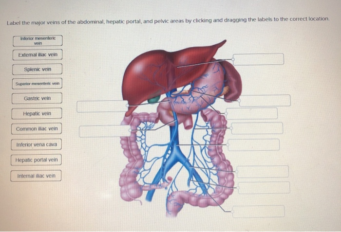
Solved Label The Major Veins Of The Abdominal Hepatic Chegg Com

Portal Hypertension Symptoms Causes And Treatment

Veins Of The Abdomen Hepatic Portal System Quiz

Anatomy Hepatic Portal Circulation Explained Youtube
Portal Venous System Wikipedia
Portal Vein Anatomy Function Embolization Thrombosis Hypertension
All About Portal Vein A Pictorial Display To Anatomy Variants And Physiopathology Insights Into Imaging Full Text

The Hepatic Portal System Is Comprised Of Veins From Almost Every Part Download Scientific Diagram

Pin On Liver Stuff

Internal Anatomy Of Liver With Branch Of Portal Vein Branch Of Hepatic Artey Template Presentation Sample Of Ppt Presentation Presentation Background Images

Increases In Hepatic Portal Vein Blood Glycemia Parallel The Capability Download Scientific Diagram

Lobules Of Liver Wikipedia
1
Hepatic Portal Vein Tributaries Portocaval Anastomoses

Hepatic Portal Veins Diagram Quizlet

Hepatic Portal System Human Anatomy Organs

Hepatic Portal System And Veins Diagram Quizlet

Right Hemiplegia And Dyspnea What Is The Link Between Cancer In Hepatic Portal Vein

Liver Segment Anatomy Portal Vein Hepatic Veins Anatomi Hand Anatomy Liver Png Pngwing
Hepatic Portal Vein Stock Illustrations 178 Hepatic Portal Vein Stock Illustrations Vectors Clipart Dreamstime
The Diagram Above Shows How Things Get To And From Class 11 Biology Cbse

9 Rat Hepatic Portal System The Pattern Of Venous Return From The Download Scientific Diagram
Portal Hypertension Practice Essentials Background Anatomy

Portal Vein An Overview Sciencedirect Topics
File 2138 Hepatic Portal Vein System Jpg Wikimedia Commons

Anatomy Of Abdominal Vessels Inews

The Hepatic Portal System Explained Youtube
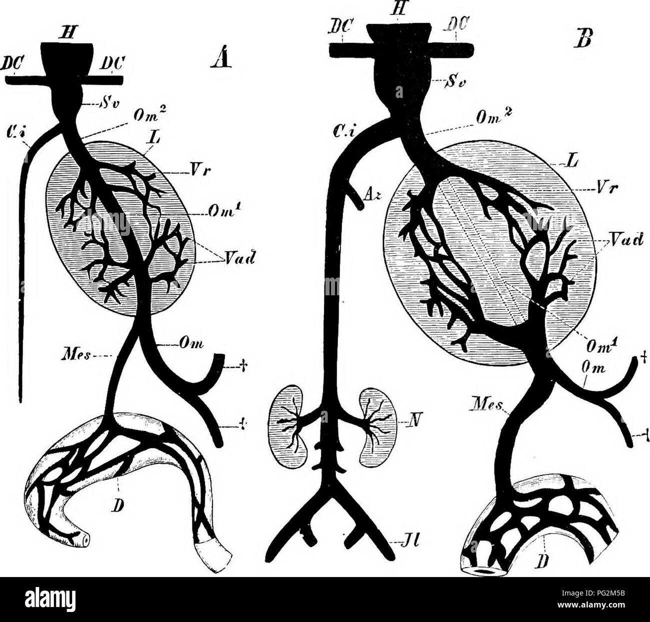
Elements Of The Comparative Anatomy Of Vertebrates Anatomy Comparative Venous System 331 A Renal Portal System Occurs In Connection With The Embryonic Kidney In All Sauropsida And Traces Of It Can

Portal Vein And Hepatic Vein In Liver
0 Response to "43 hepatic portal vein diagram"
Post a Comment