45 Skin Cross Section Diagram
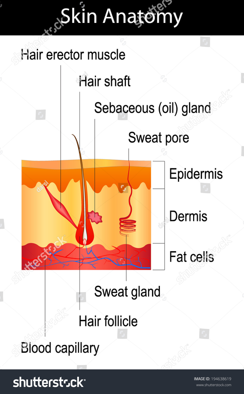
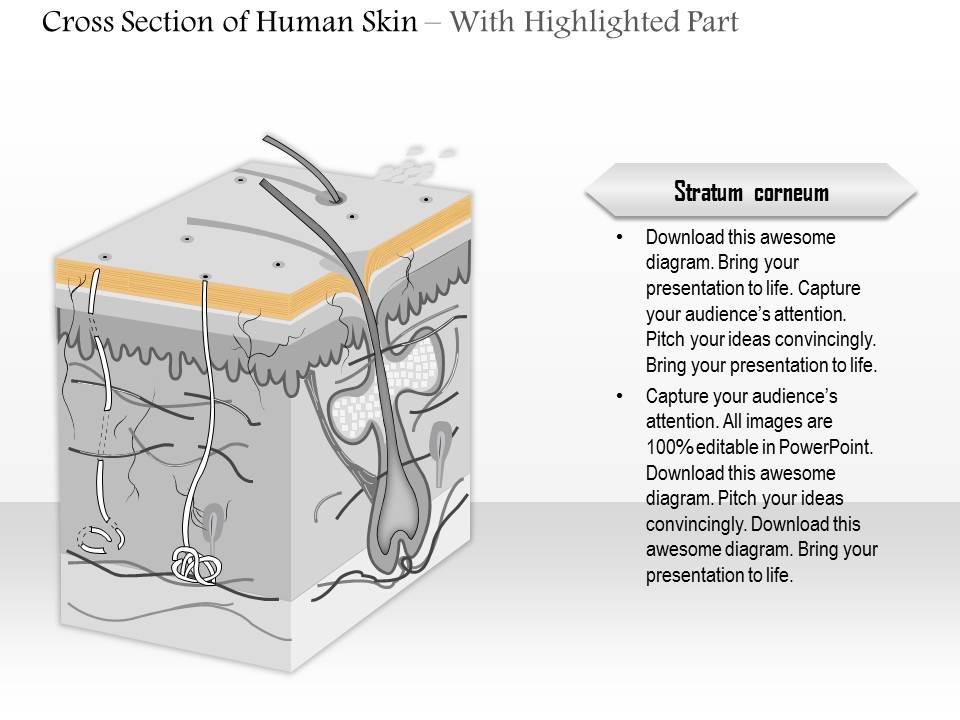
Structure of the Skin: Cross-section through the Skin ... The cross-section through the skin represents the following layers: Epidermis The epidermis is a Greek word that means 'over' or 'upon.' It is the outermost avascular (without blood vessels) layer of the skin. The epidermis consists of stratified keratinised squamous epithelium. The thickness of the epidermis varies at different body sites. Creative Diagram Of The Skin - Glaucoma Template Anatomy of skin cross section on white background. The dermis is the thicker deeper layer of the skin. Please click on the diagrams to view larger version. Subcutaneous tissue and deep fascia. This skin diagram clearly shows all the layers of skin. This is the most important function of the skin.
A Human Body Skin-structure Quiz! - ProProfs In this, a human body skin structure quiz, we are going to focus on the underlying and the most elementary structure of the human body. It's easy to take your skin for granted, but when you consider how it protects your body from harm, it is something we should appreciate more. Do you know as much as you should about it? Let's take this quiz to find out! All the best.

Skin cross section diagram
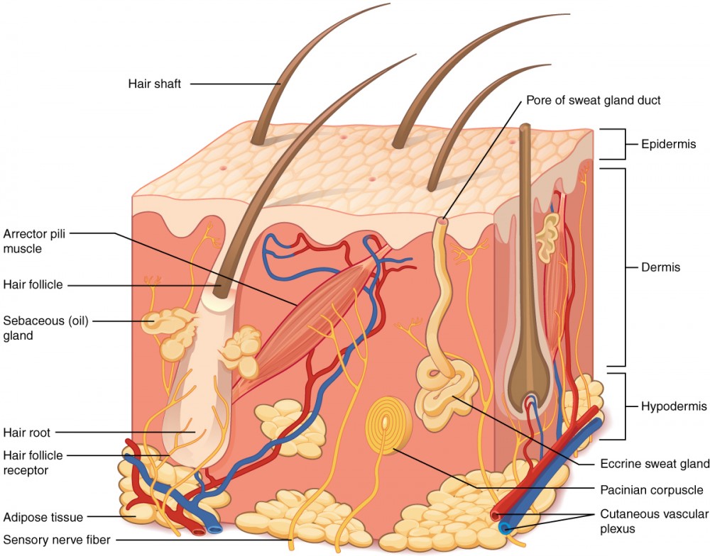
Skin: Cells, layers and histological features | Kenhub Undoubtedly, the skin is the largest organ in the human body; literally covering you from head to toe. The organ constitutes almost 8-20% of body mass and has a surface area of approximately 1.6 to 1.8 m2, in an adult. It is comprised of three major layers: epidermis, dermis and hypodermis, which contain certain sublayers. Human Body Skin Diagram - Studying Diagrams Human body parts detailed set Human body medical demographic Chart of different Human Organs Human skin detailed diagram Human skin anatomy UVB and UVA radiation Young healthy sking and older skin Skin cross-section. It shields the body against heat light injury and infection. A network of nerves transmits sensory signals to the brain. Body Cavities and Membranes: Labeled Diagram, Definitions We can use the circular cross-section below as a reference. The cross-section illustrates as if we are looking down at the spinal cord, and it shows the layers of the spinal cavity discussed above. The spinal cord shown in red is in the center of the spinal cavity. The spinal cavity is enclosed by the vertebral column shown in green.
Skin cross section diagram. Subcutaneous tissue Stock Photos and Images. 467 ... Skin subcutaneous Injection diagram Stock Photography by colematt 3 / 53 Layers Of Human Skin Pictures by PeterHermesFurian 19 / 505 cellulite cross section Stock Photos by jgroup 28 / 1,568 Epidermis of the skin Stock Photo by alila 56 / 1,794 Subcutaneous tissue Picture by radiantskies 0 / 42 Tissue repair on human skin Stock Photo by ... Phys. Rev. C 104, 024606 (2021) - Neutron skin thickness ... Neutron skin thickness of Pb 208 determined from the reaction cross section for proton scattering Shingo Tagami, Tomotsugu Wakasa, Jun Matsui, Masanobu Yahiro, and Maya Takechi Phys. Rev. C 104, 024606 - Published 9 August 2021 Structure & Function of Your Skin - American Osteopathic ... A cross-section of skin shows the major parts. It is divided into three layers. The outer layer is the epidermis. The dermis is in the middle and fat forms the innermost layer. Blood vessels, nerves, hair follicles, oil glands and sweat glands are located in the dermis. What It Does . . . Skin Cross Section Images, Stock Photos & Vectors ... 8,692 skin cross section stock photos, vectors, and illustrations are available royalty-free. See skin cross section stock video clips. of 87. scalp anatomy pigment hair skin vessels cross section of veins hair follice muscle labelled structure of the hair follicle layers epidermis hair anatomy hair section. Try these curated collections.
Can Hair Regrow After It Falls Out? 2022 - Hair Loss Geeks Female pattern baldness set with skin cross-section diagram. Treatment result in top view. Alopecia infographics vector template for clinics. Page Contents. What is Hair Loss? ... Lichen planopilaris, for starters, is one skin condition that causes dry, flaky rashes, sometimes with redness. It causes hair to come out in clumps and affects more ... Cross sectional anatomy - Kenhub Cross section through the tongue and C2: Diagram This cross-section has the exact same orientation as the previous one. The posterior landmark is provided by the second cervical vertebra (axis) while the anterior one is provided by the tongue. However, there are quite a few differences between them. Why testicles have tastebuds: Viral TikTok exposes a weird ... A cross-section diagram of a taste bud on a human tongue. ... Application of food to scrotal skin has no effect, but science shows the testis can be sensitive to the ingestion of certain foods. Body Planes and Sections: Anatomical Position, Directional ... Anatomical position, body planes, anatomy sections. Directional term descriptions, definitions, example labeled diagrams of sagittal, coronal, transverse, oblique, and longitudinal axis. Quiz yourself on how each plane divides the body into front (anterior) and back (posterior) portions, right and l
Integumentary System Worksheet Basic Skin Structure Exam Instructions. Skin Cross Section. a synapse D. Get straight to the crux of the matter with this worksheet comprising diagrams of the six organ systems. Cpt codes list excel A cell is the smallest and most basic form of life. Robert Hooke, one of the first scientists to use a light microscope, discovered the cell in 1665. Human Skin Cross Section Images, Stock Photos & Vectors ... Find human skin cross section stock images in HD and millions of other royalty-free stock photos, illustrations and vectors in the Shutterstock collection. Thousands of new, high-quality pictures added every day. What Is the Relationship between the Skin and Homeostasis? A person sweating when they are hot is an example of the skin helping to regulate body temperature. During temperature homeostasis, or thermoregulation, the skin and homeostasis cause the body to sweat.When the skin senses that the body is heating up because of the environment's temperature, the hypothalamus sends a signal via nerves to sweat glands and blood vessels in the skin. Skin Anatomy: The Layers of Skin and Their Functions The epidermis is made up of five individual layers: 2. Stratum basale: This bottom layer, also known as the basal cell layer, has column-shaped cells that push older cells toward the surface. As the cells move upward, they start to flatten and die. The layer is also made up of melanocytes (that produce a pigment that gives the skin its color ...
Aging Skin. Cross Section Young And Old Skin. The Diagram ...
Bowen's Disease. Symptoms and treatment of Bowen ... - Patient Skin diagram cross-section view The epidermis has three main types of cell: Basal cells. These are the bottom layer of cells in the epidermis. Keratinocytes. These cells are in layers above the basal layer. They make a substance called keratin which is a hard waxy material.
Human Skin Crosssection Hair Structure Diagram Stock Vector ...
Anatomy, Skin (Integument), Epidermis - StatPearls - NCBI ... Skin is the largest organ in the body and covers the body's entire external surface. It is made up of three layers, the epidermis, dermis, and the hypodermis, all three of which vary significantly in their anatomy and function. The skin's structure is made up of an intricate network which serves as the body's initial barrier against pathogens, UV light, and chemicals, and mechanical injury.
skin, skinned, cross-section diagram - Stock Illustration ...
Integumentary Layers Of Skin Structure - about the ... Integumentary Layers Of Skin Structure. Here are a number of highest rated Integumentary Layers Of Skin Structure pictures on internet. We identified it from well-behaved source. Its submitted by management in the best field. We assume this kind of Integumentary Layers Of Skin Structure graphic could possibly be the most trending topic with we ...
Skin Cross-Section - Anatomy and Physiology
Integumentary System - Innerbody The skin is only a few millimeters thick yet is by far the largest organ in the body. The average person's skin weighs 10 pounds and has a surface area of almost 20 square feet. Skin forms the body's outer covering and forms a barrier to protect the body from chemicals, disease, UV light, and physical damage.
Layers of the Skin | Anatomy and Physiology I
Cross Section Diagram - structure of a volcano youtube ... Cross Section Diagram. Here are a number of highest rated Cross Section Diagram pictures upon internet. We identified it from well-behaved source. Its submitted by giving out in the best field. We take this kind of Cross Section Diagram graphic could possibly be the most trending subject next we portion it in google plus or facebook.

Skin cross section Pictures, Skin cross section Stock Photos ...
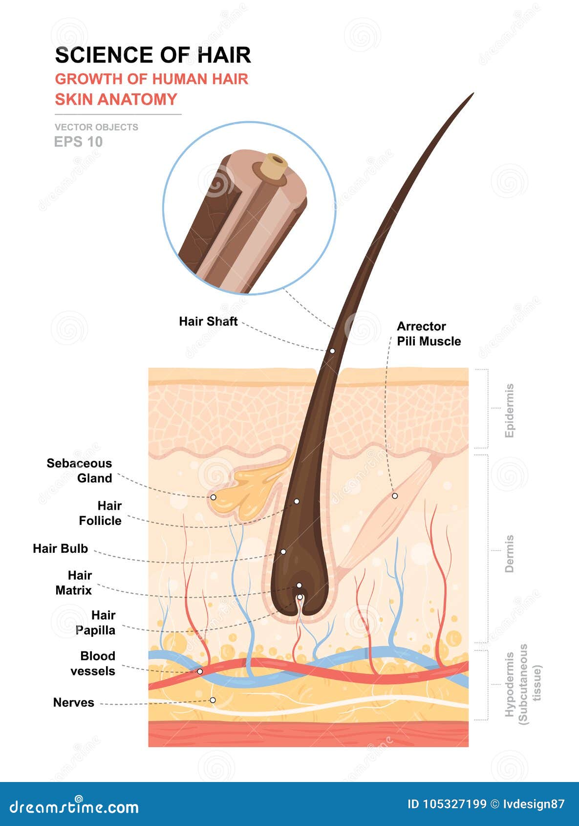
Anatomy, Hair Follicle - StatPearls - NCBI Bookshelf Cross section of layers of the skin. Hair follicles, hair roots and hair shafts, sweat glands, pores, epidermis, dermis, hypodermis. Papillary and reticular layer. Eccrine sweat gland. Arrector pili muscles, sebaceous oil glands. Contributed by Chelsea
Human Skin Cross Section Stock Illustrations – 759 Human Skin ...
Wing Geometry Definitions - NASA The total surface area includes both upper and lower surfaces. The wing area is a projected area and is almost half of the total surface area. The aspect ratio (AR) of a wing is defined to be the square of the span (s) divided by the wing area (A). Aspect ratio is a measure of how long and slender a wing is from tip to tip.

Human Skin Cell Crosssection Of The Structure Labeled Hair ...
Musculoskeletal System - Skeletal System: TEAS ... Diagram of cross-section of cortical bone showing osteocytes and osteon. Cortical bone, which is the stronger of these two types of bone, is the kind of bone that is most needed for bodily support and bodily movement. ... a closed fracture is defined as one that does not break through the surface of the skin and this type of fracture and this ...

Skin Anatomy. Cross Section Of The Human Skin. Layers Of The ...
Pore Illustrations and Clip Art. 2,904 Pore royalty free ... Vector Stock Illustration by emaria 15 / 1,133 Human skin anatomy Stock Illustration by bluering 17 / 196 Human skin section diagram Stock Illustrations by Pixelchaos 61 / 2,947 A typical animal cell, eps10 Stock Illustration by alila 12 / 1,186 Blackhead Drawings by rob3000 5 / 135 Cell structure Stock Illustrations by megija 5 / 202 Alveoli ...

Skin - Visual Dictionary
Skin and skin appendage - Knowledge @ AMBOSS The skin is the largest organ of the body, covering an area of approximately 2 m 2.The skin is composed of the cutis (including the dermis and epidermis), subcutaneous tissue, and skin appendages.The epidermis, which is derived from ectoderm, is the outermost layer of the skin and is mainly composed of keratinocytes.The dermis, which is derived from mesoderm, is located underneath the ...

Free art print of Human skin anatomy
Wire Gauge and Current Limits Including Skin ... - PowerStream Wire cross section in circular mils This chart of American Wire Gauge (AWG) wire sizes and rated ampacities is data intended for the pleasure of our readers only. Typographical errors, etc. are probable, since the typist is not a professional (our CEO).

Skin cross section art print poster
Body Cavities and Membranes: Labeled Diagram, Definitions We can use the circular cross-section below as a reference. The cross-section illustrates as if we are looking down at the spinal cord, and it shows the layers of the spinal cavity discussed above. The spinal cord shown in red is in the center of the spinal cavity. The spinal cavity is enclosed by the vertebral column shown in green.

Chapter 1: Skin Cross Section Diagram | Quizlet
Human Body Skin Diagram - Studying Diagrams Human body parts detailed set Human body medical demographic Chart of different Human Organs Human skin detailed diagram Human skin anatomy UVB and UVA radiation Young healthy sking and older skin Skin cross-section. It shields the body against heat light injury and infection. A network of nerves transmits sensory signals to the brain.

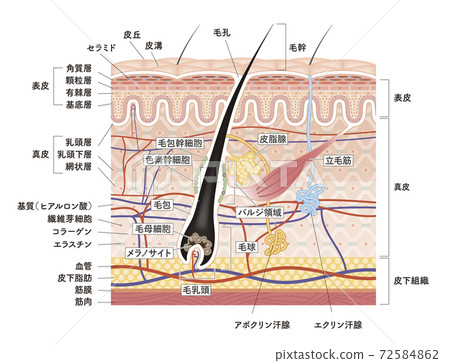
Human Skin And Hair Structure. Cross Section Of The Human ...
Skin: Cells, layers and histological features | Kenhub Undoubtedly, the skin is the largest organ in the human body; literally covering you from head to toe. The organ constitutes almost 8-20% of body mass and has a surface area of approximately 1.6 to 1.8 m2, in an adult. It is comprised of three major layers: epidermis, dermis and hypodermis, which contain certain sublayers.

Human skin anatomy. Human skin cross section, eps8. | CanStock
0614 cross section of human skin Medical Images For ...

Cross Section Human Skin. Royalty Free Cliparts, Vectors, And ...

Skin Cross-Section Integumentary System Diagram | Quizlet

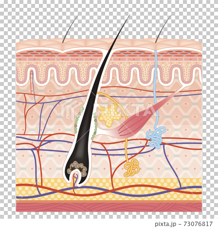
Diagram of a hair follicle in a cross section of skin layers
![A schematic cross-section of human skin [9]. | Download ...](https://www.researchgate.net/publication/269774030/figure/fig1/AS:601641325715472@1520453878744/A-schematic-cross-section-of-human-skin-9_Q640.jpg)
A schematic cross-section of human skin [9]. | Download ...

✓ Human Skin Cross-Section vector illustration of the ...
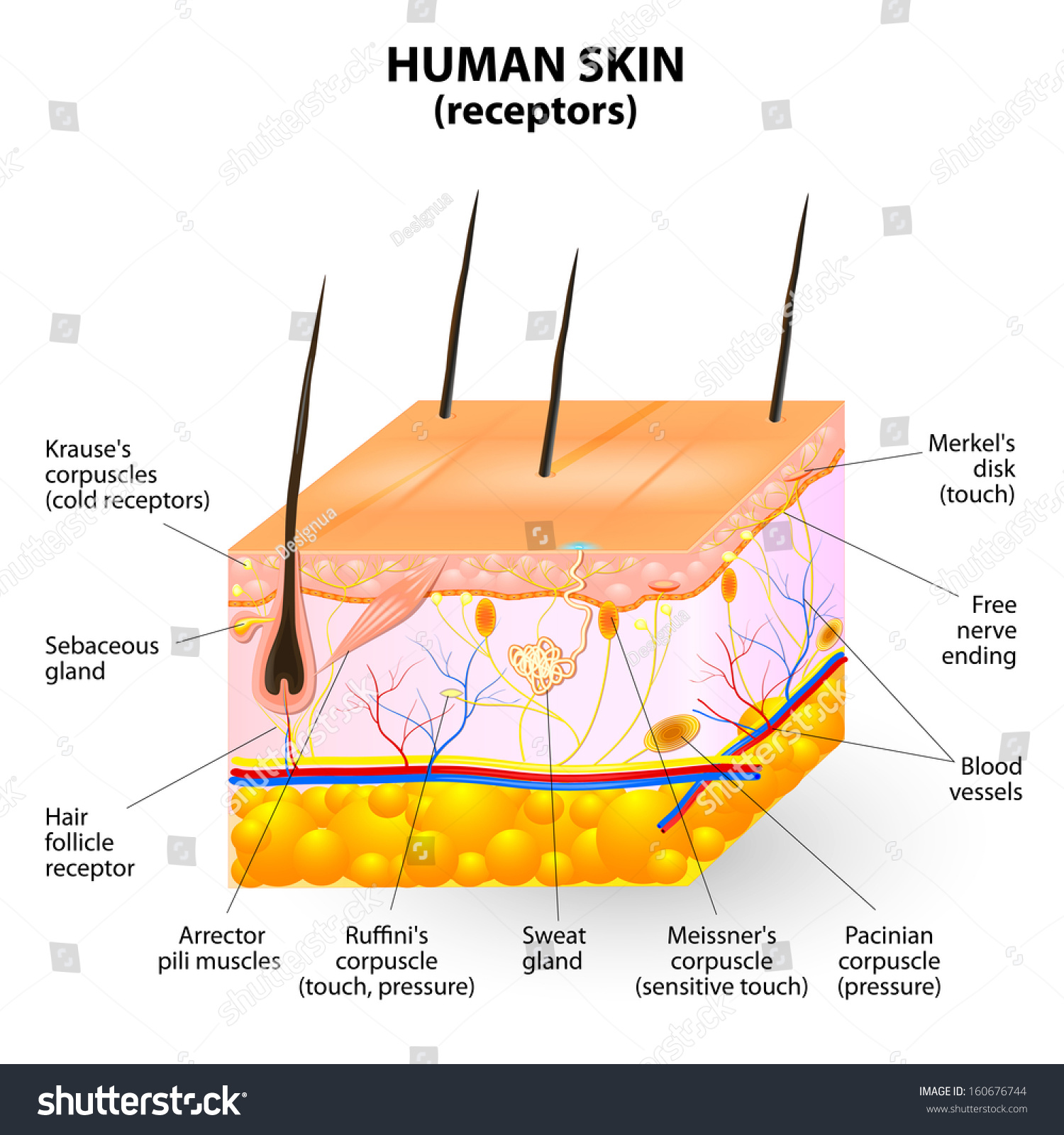
cross section human skin. Pressure, vibration, - Royalty Free ...

Perspiring anatomical skin cross section vector illustration ...

Skin Cross Section Diagram Vector Images (over 130)

Cross section of skin — Science Learning Hub

Cross-sectional view of skin | Download Scientific Diagram
![Figure, Cross section of layers of...] - StatPearls - NCBI ...](https://www.ncbi.nlm.nih.gov/books/NBK470464/bin/skin__layers_hair__follicles_sweat__glands.jpg)
Figure, Cross section of layers of...] - StatPearls - NCBI ...

1 The cross section of skin shows the three top main layers ...

4-A cross-sectional representation of the anatomy of human ...
skin, cross-section diagram, vector - Stock Illustration ...
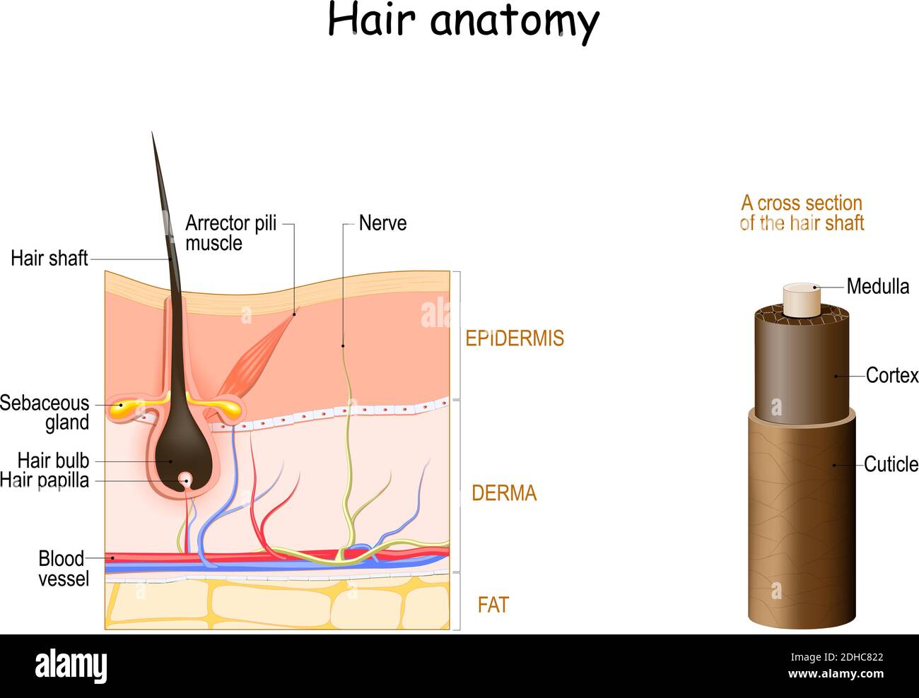
Hair anatomy. Cross section of the hair shaft. skin layers ...
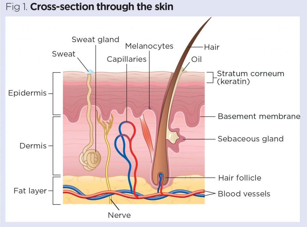
Skin 1: the structure and functions of the skin | Nursing Times

Cross section of the skin. | Download Scientific Diagram
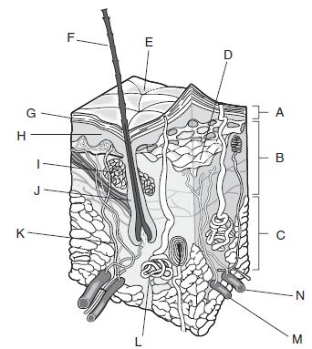
Label the following diagram of a cross-section of the skin ...

Cross section of skin. 26 | Download Scientific Diagram

Anatomy of the skin Brownsville OBGYN Associates | Texas
Illustration of skin cross section on white background ...
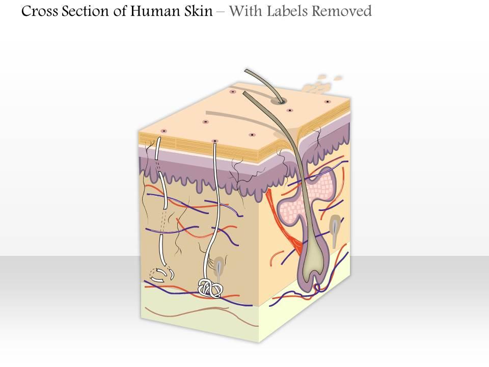
0614 cross section of human skin Medical Images For ...

Skin Cross Section Diagram | Quizlet

Drawings - Human skin section diagram. Stock Illustration ...
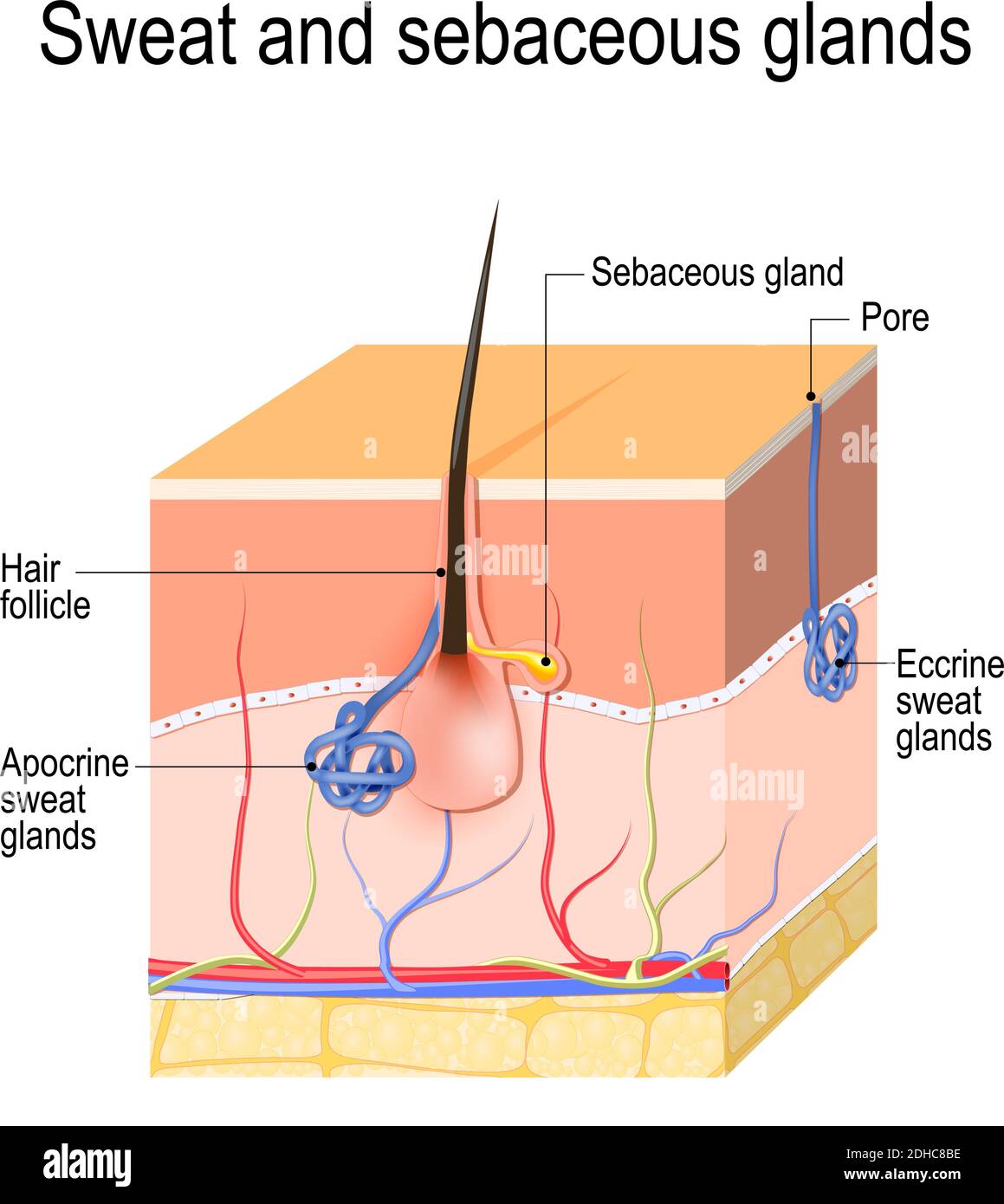
Sweat glands (apocrine, eccrine) and sebaceous gland. Cross ...

8: Schematic cross section of the human skin. (©The McGraw ...

Skin Anatomy - EnchantedLearning.com | Skin anatomy, Skin ...

76 Skin Cross Section Diagram Drawing Illustrations & Clip ...

Skin Crosssection Stock Photo - Download Image Now - iStock
0 Response to "45 Skin Cross Section Diagram"
Post a Comment